*Important notice: This news reports on an unedited version of an accepted paper and is awaiting final editing. Therefore, the paper should not be regarded as conclusive or treated as established information.
A label-free imaging method has tracked how human lung cancer cells respond to X-ray radiation. Combining stimulated Raman scattering (SRS) with two-photon excitation fluorescence (TPEF) microscopy, the approach monitors changes in lipids, proteins, and mitochondrial oxidative stress without chemical dyes, detecting cellular changes at doses as low as 2 gray (Gy). These findings were published in Scientific Reports.
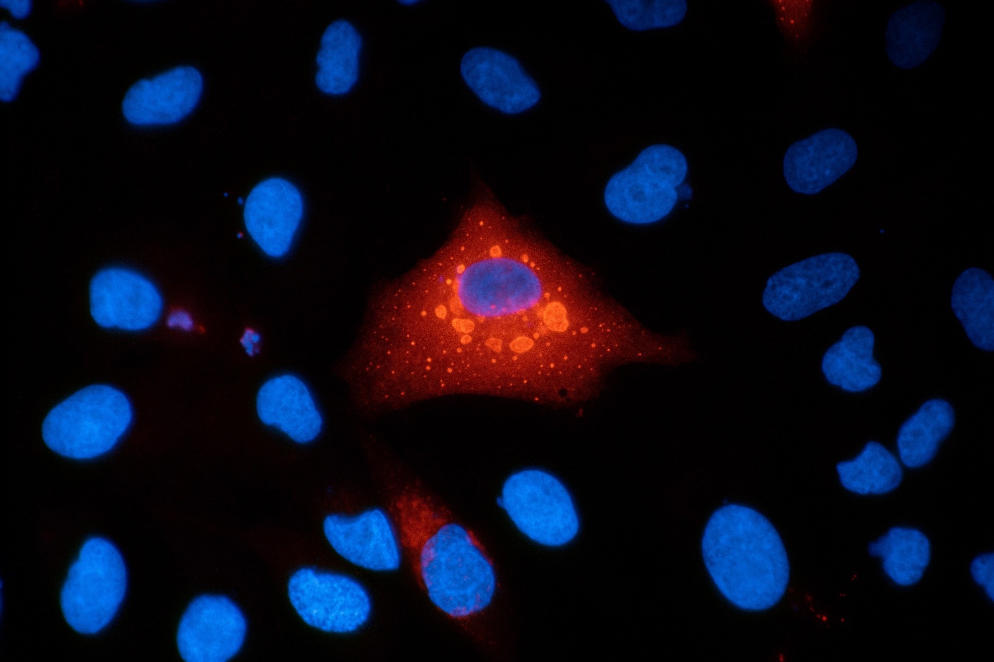 Study: Label-free multimodal nonlinear optical imaging reveals key biomarkers of cellular radiation response. Image Credit: VANOXmicroscopy/Shutterstock.com
Why Radiation Research Needs Better Cell Imaging
Radiation damage is commonly assessed using chromosome damage tests and cell survival assays, which often require chemical fixation or fluorescent dyes. However, these methods can obscure natural changes over time, as dyes can affect cell metabolism.
In contrast, nonlinear optical microscopy allows for the investigation of cells without added dyes. SRS detects molecular vibrations to map cellular components, while TPEF captures signals from metabolic cofactors like reduced nicotinamide adenine dinucleotide phosphate [NAD(P)H] and flavin adenine dinucleotide (FAD). Combining these techniques enables researchers to track dynamic changes in cell chemistry over time while keeping cells intact.
Experimental Design and Imaging Methodology
To investigate cellular radiobiology under clinical conditions, researchers cultured human lung adenocarcinoma cells (A549) on glass-bottom dishes and exposed them to X-ray doses ranging from 2 to 10 Gy at a rate of 1 Gy per minute. The experiment utilized 120 kVp X-rays, with sham-irradiated controls maintained under identical conditions. After exposure, the cells were returned to incubators and imaged systematically at 24, 48, and 72 hours.
Download a free copy of this page here!
The imaging system combined spectral-focusing SRS microscopy with TPEF. An optical parametric oscillator supplied a fixed 1040 nm Stokes pulse and a tunable pump beam at an 80 MHz repetition rate. High-refractive-index glass blocks stretched the pulses to resolve molecular vibrations associated with lipids at 2850 cm-1 and proteins at 2926 cm-1.
Raman images were analyzed using non-negative least-squares spectral unmixing and calibrated against oleic acid and bovine serum albumin standards. MATLAB software was employed to identify cell boundaries and mitochondria-rich regions.
Across 18 fields of view for each dose and time point, with triplicate runs, the study measured cell size, biochemical composition, and organelle distribution. Area-normalized measurements helped separate changes in cellular composition from differences in cell size or density.
X-Ray Exposure Triggers Changes in Cancer Cell Lipids and Metabolism
The imaging showed dose- and time-dependent changes in cell size, lipid content, protein levels, and mitochondrial redox state. Irradiated cells
grew larger and accumulated more lipid droplets; at 48 and 72 hours, the droplets expanded faster than the surrounding cells. This pattern may reflect radiation-induced senescence and cellular stress.
Lipid content increased while area-normalized protein content declined, particularly at 48 hours. As a result, the lipid-to-protein ratio increased with radiation dose across the observation period. The optical redox ratio (ORR) increased significantly at 48 and 72 hours, primarily due to a decrease in NAD(P)H at 48 hours. By 72 hours, increased FAD contributed more to this shift, indicating a change in cellular redox state.
The study also indicated increased spatial overlap between lipid droplets and FAD-rich mitochondria over 72 hours, with no clear fragmentation of the mitochondrial network observed. This close association may reflect changes in how cells manage lipids during radiation-induced stress, although the imaging data alone cannot establish the mechanisms.
Applications for Cancer Treatment and Radiation Safety
This label-free imaging method has significant potential for cancer therapy and occupational radiation safety. In cancer treatment, it could help assess tumor responses to radiation using biopsy samples or organoids, potentially providing earlier information than conventional cell survival tests, which can take weeks.
Furthermore, the method could support radiation exposure assessments in workplaces and emergency settings. After accidental exposure, changes in the lipid-to-protein ratio, lipid droplet size, and cellular redox state could help evaluate radiation-induced cell damage without prolonged cell culture. However, further real-world testing is needed to determine whether these markers can reliably estimate radiation doses in exposed individuals.
Toward More Sensitive Detection of Radiation-Induced Cell Changes
Through a method that combines Raman scattering and multiphoton autofluorescence, researchers tracked radiation-induced changes in living cancer cells without chemical dyes by detecting alterations in cellular lipids, proteins, and redox state after X-ray exposure.
Future work could involve testing these optical markers in healthy cells, including lung fibroblasts and white blood cells, to compare their responses with those of cancer cells. Machine learning (ML) algorithms could automate cell and organelle tracking, enabling the detection of subtle changes at lower radiation doses.
Ultimately, though more validation is necessary, the method is a promising novel approach to detect changes in cells after X-ray exposure.
Journal Reference
Gagnon, J.R., et al. (2026). Label-free multimodal nonlinear optical imaging reveals key biomarkers of cellular radiation response. Scientific Reports. DOI: 10.1038/s41598-026-73913-w, https://www.nature.com/articles/s41598-026-73913-w.
Disclaimer: The views expressed here are those of the author expressed in their private capacity and do not necessarily represent the views of AZoM.com Limited T/A AZoNetwork the owner and operator of this website. This disclaimer forms part of the Terms and conditions of use of this website.