Microplastic (MP) monitoring in marine surface waters is often analytically challenging. This is because particle counts can fluctuate substantially and particles are embedded in complex mixes of biological, mineral, and other non-plastic materials.
Reliable characterization therefore necessitates chemical identification rather than visual classification alone, especially for microscopic particles that are difficult to identify by shape. FTIR imaging solves this problem by creating spatially detailed chemical maps of whole filter surfaces.
The particles are recognized and identified simply based on their spectrum, allowing for polymer-specific identification while also providing particle-level size and morphology data from the same experiment.
This article outlines how Dr. J. Paulo Felizardo and Dr. C. Rodrigues from UFF Brazil used FTIR imaging to characterize microplastics in Southern Ocean seawater samples obtained during the International Antarctic Coastal Circumnavigation Expedition (2024–2025).
Experimental
Surface water samples were obtained at various points throughout the Southern Ocean. At each station, 3 L of seawater was extracted using a Niskin bottle and promptly vacuum-filtered in situ through pre-muffled stainless steel meshes with a pore size of 25 µm.
The loaded filters were then chilled and brought to the laboratory, where they were used directly in the extraction technique outlined below.
For sample preparation, the filters were processed sequentially. First, organic matter was digested with a 10% KOH solution at a 10:1 ratio to the sample volume, then incubated at 40 °C for 72 hours.
The samples were refiltered using a stainless steel mesh. For density separation, each filter received 10–15 mL of NaI solution (4.4 mol/L; 1.8 g/cm3). The samples were then sonicated for five minutes at 50 kHz, magnetically swirled for five minutes, and centrifuged at 7000 rpm for two minutes.
The supernatant was then vacuum-filtered through 0.2 µm aluminum oxide (Anodisc) filters. To verify the sample preparation method, a procedural blank was run through all processes except the ambient water.
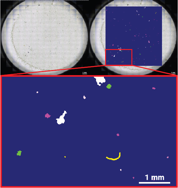
Fig 1. Exemplary data obtained from a Southern Ocean surface seawater sample. Left: transmitted-light mosaic of the Anodisc filter. Right: corresponding hyperspectral μ-FTIR image acquired with the Bruker LUMOS II and evaluated with the Bruker MPID software. Image Credit: Dr. J. Paulo Felizardo and Dr. C. Rodrigues
Results
Three Southern Ocean samples were analyzed, and microplastic concentrations ranged between 9000 and 32,000 MPs/m3. Published results for Southern Ocean surface waters range from 0.01 to 15,400 MPs per m3, indicating significant methodological heterogeneity between research.
The higher concentrations shown here are likely due to the analytical workflow's lower effective size threshold, which captures microscopic particles that are not retained or reported in many traditional studies employing non-imaging methodologies or subsampling techniques.
Polymer composition varied among sampling stations. Polypropylene predominated at most sites, accounting for 68–81% of detected particles. Poly(ethylene terephthalate) achieved a relative abundance of about 52% at one location.
The average particle size ranged from 40 to 160 µm depending on the polymer type. The procedural blank had a very low particle level, indicating that there was negligible background contamination from laboratory handling.
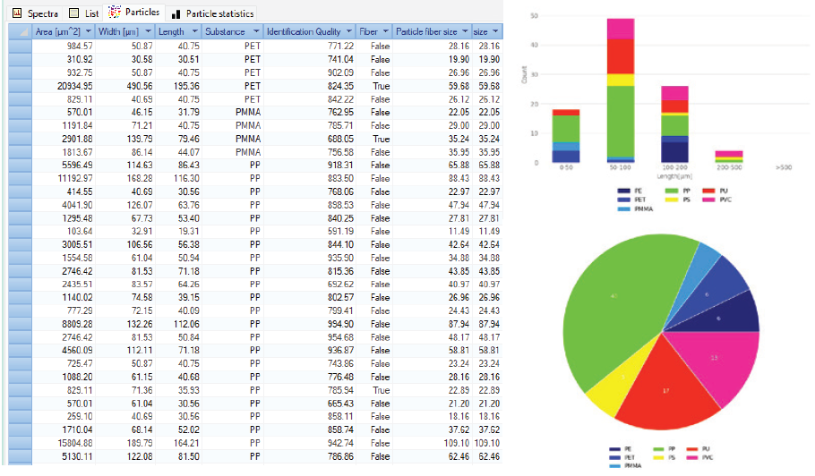
Fig 2. Exemplary MPID result obtained from a Southern Ocean subsurface seawater sample. Extensive particle information is given together with an interactive classification map. When a particle or particle class is selected, all respective information is directly displayed. Image Credit: Bruker Optics
Conclusion
The LUMOS II FTIR imaging microscope, paired with MPID automated processing, allows for automated particle detection and polymer-specific identification on Anodisc filters while maintaining particle-level size and morphology data.
The equipment detects and chemically identifies particles from approximately 10 µm upward, providing concentration estimations that include the small-particle percentage available for µ-FTIR imaging.
The effectiveness of MPID, software that automatically evaluates entire FTIR spectra of microplastic samples in a matter of minutes, was demonstrated. It produced detailed statistics for all particles on the filter and classified them by number, identity, and size.
The Southern Ocean samples included 9000 to 32,000 MPs/m3. These concentrations are not directly comparable to levels obtained with less sensitive sampling techniques, such as 300 µm mesh nets.
As such approaches exclude much of the small-particle fraction found by FTIR imaging, the higher amounts reported here are primarily due to changes in the observable particle-size range and should not be regarded as direct evidence of increased environmental contamination.
The concentrations were computed from 3 L samples and extrapolated to 1 m3, indicating that minor changes in particle counts can significantly impact the extrapolated concentration. The small sample volume may not adequately capture regional variability in particle abundance.
These considerations should be addressed when interpreting the findings and comparing them to other investigations. Harmonized sampling and analytical techniques, such as uniform particle-size ranges and IR imaging-based identification, are thus necessary to improve comparability across studies.
Acknowledgments
Produced using materials originally authored by João Paulo Felizardo (PhD), Geochemistry Program, Federal Fluminense University; Camila Rodrigues e Silva (PhD), Geochemistry Program, Federal Fluminense University; and Alexander Staub (PhD), Product Manager IR Microscopy, Bruker Optics GmbH & Co. KG, Ettlingen.

This information has been sourced, reviewed, and adapted from materials provided by Bruker Optics.
For more information on this source, please visit Bruker Optics.