Mussels are well-known sentinel organisms for detecting coastal microplastics. As suspension feeders, they filter large volumes of water and collect particles from the surrounding water column, and their tissue load reflects local pollution over time.
To determine the polymer identification of the accumulating particles, chemical analysis is required rather than visual inspection alone. Particles recovered from digested tissue vary in size and coexist with mineral and organic waste that cannot be separated visually from plastics.
FTIR imaging can generate spatially detailed chemical maps of large filter regions and even complete filters, identifying each particle by its infrared spectrum and providing size and morphological data in the same measurement.
This article discusses its use for microplastic characterization in two mussel species from Guanabara Bay, Rio de Janeiro, sampled across two seasons, as described by Doctors J. Paulo Felizardo and C. Rodrigues of UFF, Brazil.
Experimental
In total, 120 mussels were taken from two aquaculture locations in Guanabara Bay: Jurujuba and the Rio-Niterói Bridge area.
Two species were examined: the brown mussel, Perna perna, and the Asian green mussel, Perna viridis. Collections were conducted during the rainy season (February 2025) and the dry season (August 2025) to account for seasonal fluctuation in particle burden.
Sample preparation was performed by alkaline digestion with a 10% KOH solution at a solution-to-sample volume ratio of 5:1. Samples were incubated at 40 °C for 72 hours. The digested material was filtered through a 25 µm stainless steel mesh, and the remaining portion was put into glass beakers.
To separate the density, 10 mL of NaI solution (4.4 mol/L; 1.8 g/cm3) was added, followed by 15 minutes of sonication and mechanical agitation. During agitation, another 10 mL of NaI solution was added.
The cycle was repeated twice. The samples were centrifuged at 500 × g for about two minutes, and repeated if any suspended material remained.
The low-density supernatant was vacuum-filtered using 0.2 µm Anodisc aluminum oxide filters. To validate the sample preparation method, a procedural blank was run through all phases while excluding the biological tissue.
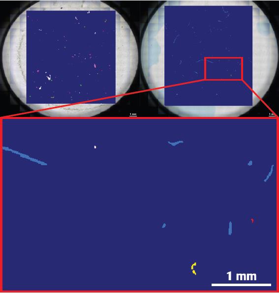
Fig 1. Data obtained from a Guanabara Bay mussel tissue sample. Left: transmitted-light mosaic of the Anodisc filter following alkaline digestion and density-based extraction. Right: corresponding hyperspectral μFT-IR map acquired with the Bruker LUMOS II and evaluated with the Bruker MPID software. Image Credit: Dr. J. Paulo Felizardo and Dr. C.Rodrigues
Results
Anodisc filters were studied in transmittance mode using the Bruker LUMOS II FT-IR imaging microscope, and data was processed using the Microplastic Identifier (MPID) software, which detects and identifies particles using machine learning.
Microplastic particles were detected in all 120 sampled mussels within the investigated size range, demonstrating broad exposure in Guanabara Bay using the applicable preparation and µFTIR imaging workflow. This 100% occurrence rate reflects the severity of particulate pollution in Guanabara Bay, as well as the analytical method's sensitivity.
The data revealed that Perna viridis had a greater absolute particle count per individual (mean 5.4 particles) than P. perna (mean 2.2 particles).
When normalized to soft tissue mass, the relationship reversed: P. perna had considerably higher contamination (0.54 ± 0.45 particles/g) than P. viridis (0.19 ± 0.13).
This difference may represent species-specific body mass, tissue composition, filtration behavior, or site-specific exposure patterns, and should thus be interpreted in light of biological and environmental factors.
Polypropylene (PP) was the major synthetic polymer found in both mussel species. In P. perna, PP accounted for 92% of fragments, with mean dimensions of 4.33 µm width and 35.77 µm length.
Its frequency is consistent with polypropylene's low density and widespread use in packaging, ropes, and other marine-related goods, although the polymer's identity alone does not allow source identification.
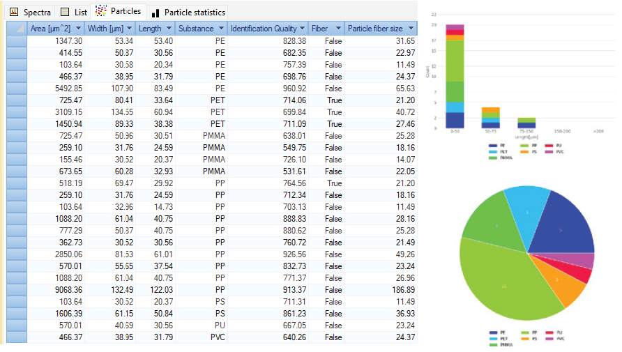
Fig 2. Exemplary MPID result obtained from a Guanabara Bay mussel tissue sample. Extensive particle information is given together with an interactive classification map. When a particle or particle class is selected, all respective information is directly displayed. Image Credit: Bruker Optics
Conclusion
FTIR microspectroscopy is ideal for analyzing microplastics in digested biological tissues because it detects particles smaller than the visual-sorting size limit and allows for automated, chemically specific polymer identification, reducing operator variability.
Microplastics in all mussel samples indicate the magnitude of contamination in Guanabara Bay. The differences in mass-normalized loads between P. perna and P. viridis suggest that species-specific biological parameters should be included in conventional biomonitoring techniques.
Mussels, as filter feeders, gather microplastics from the surrounding water and can be used as bioindicators of environmental contamination. Their presence in marine food chains and consumption as seafood provide a potential exposure pathway for higher trophic levels. Continuous monitoring is therefore required to determine environmental contamination and potential human exposure.
Acknowledgments
Produced using materials originally authored by João Paulo Felizardo (PhD), Geochemistry Program, Federal Fluminense University; Camila Rodrigues e Silva (PhD), Geochemistry Program, Federal Fluminense University; Alexander Staub (PhD), Product Manager, IR Microscopy, Bruker Optics GmbH & Co. KG.

This information has been sourced, reviewed, and adapted from materials provided by Bruker Optics.
For more information on this source, please visit Bruker Optics.