Can microplastics pass through biological barriers and accumulate inside organs? This has emerged as one of the most pressing questions in environmental and biological research.
While the presence of microplastics in digestive systems is well documented, direct detection within organs remains analytically difficult. To investigate this, a university research group looked at the possibility that sufficiently small polyethylene (PE) microplastics could breach the intestinal barrier and accumulate in liver tissue.
This article details the analytical method used to obtain chemically specific evidence of PE particles in the tissue. Cryo-sectioned chicken liver samples were used as a model system. Under optical microscopy, tiny black inclusions were visible within the tissue (Figure 1).
Because shape and optical contrast alone were insufficient to detect their chemical makeup, Raman microscopy was utilized to separate putative PE particles from biological structures.
Raman Microscopy for the Smallest Particles
Despite being the established standard, infrared-based research did not give conclusive evidence of polyethylene in liver tissue; infrared microscopy had difficulty resolving particles of 1–5 µm.
Raman microscopy was chosen because of its excellent spatial resolution, which allows the chemical identification of particles in this size range. This property makes Raman spectroscopy ideal for detecting very small microplastic particles in biological tissue.
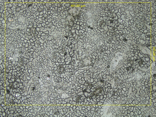
Fig 1. Microscopic image of a chicken liver cryo-section. Dark spots are suspected to be microplastic particles. Image Credit: Bruker Optics
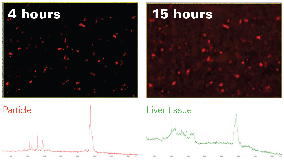
Fig 2. Comparison of the Raman image acquired with Random Scanning after four hours (top left) with the complete 15-hour Raman image (top right). Clearly, most particles have already been found by the algorithm. Found particles were unambiguously identified as PE (bottom left) and clearly stand out from the liver tissue matrix (bottom right). Image Credit: Bruker Optics
Results
The mapping area was roughly 491 × 345 µm, with a spatial sampling resolution of 1.2 µm. This resulted in a dataset that can resolve micrometer-scale inclusions inside the tissue matrix.
To achieve complete coverage, a standard grid-based Raman mapping was expected to take 15 hours in total (Figure 2, top right). However, because the target particles were predicted to be sparse and dispersed unevenly, Random Scanning was used to avoid devoting measurement time to tissue regions with no meaningful signals.
This method increased the likelihood of finding polyethylene particles by prioritizing sampling chemically informative places within the targeted area while shortening the time required to obtain relevant results.
During the first three to four hours, the system had identified and characterized most of the polyethylene particles (Figure 2, upper left). Even at this intermediate stage, the generated Raman image was spatially and statistically similar to the final dataset.
Each highlighted pixel included the distinctive Raman signature of polyethylene. The related full-range spectra (Figure 2, bottom) corroborated the chemical identity of the particles, providing clear evidence of polyethylene microplastics in liver tissue.
Conclusion
This study successfully detected and identified polyethylene microplastics in liver tissue using confocal Raman microscopy and Random Scanning.
Raman microscopy proved to be very useful for analyzing minute particles in complex biological matrices that require high spatial resolution. As a result, it serves as an effective supplement to infrared technologies for research that goes beyond their resolution limitations.
Infrared imaging, on the other hand, is a key component of microplastics investigation due to its unparalleled high-throughput screening capabilities and measurement speed.
Acknowledgments
Produced using materials originally authored by Dr. Yan Di, Product Manager, Raman Microscopy, Bruker Optics GmbH & Co. KG.

This information has been sourced, reviewed, and adapted from materials provided by Bruker Optics.
For more information on this source, please visit Bruker Optics.