In a recent article published in the journal Light: Advanced Manufacturing, researchers presented a comprehensive analysis of optical percutaneous needle biopsy (PNB), an emerging technology that integrates standard tissue sampling with advanced optical spectroscopy and imaging techniques to enhance tumor diagnostics by enabling real-time optical assessment of tissue at the needle tip during biopsy procedures.
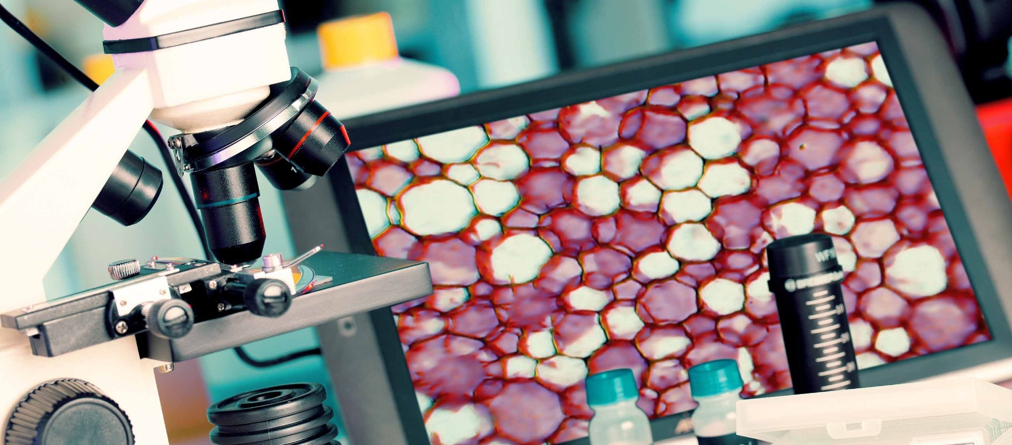
Image Credit: luchschenF/Shutterstock.com
Background
Image-guided PNB is widely regarded as the standard approach for evaluating suspected cancers. It plays a critical role in determining disease stage and identifying primary or metastatic sites. Definitive confirmation of tumor pathology depends on subsequent histological and cytological analysis.
PNB can be performed either as a core (trephine) biopsy or as a fine-needle aspiration biopsy. While fine-needle aspiration reduces tissue trauma, trephine biopsy provides larger, higher-quality histological specimens. These more substantial samples are essential for obtaining detailed insight into the carcinogenic process, including assessment of tumor invasion.
Advances in fibre optics, light sources, and detectors have facilitated the integration of optical methods into image-guided PNB. These methods include Optical Coherence Tomography (OCT), which provides high-resolution imaging of tissue morphology at the microscopic level, aiding in visualizing pathological tissue and identifying damaged vessels. Raman Spectroscopy (RS) is utilized to study the molecular signatures of tissues, guiding surgical tools for targeted brain neoplasm biopsies. Laser Doppler Flowmetry (LDF) dynamically assesses microvascular perfusion by measuring the Doppler shift from moving red blood cells.
Studies Highlighted in the Review
The review concentrates on Diffuse Reflectance Spectroscopy (DRS) and Fluorescence Spectroscopy (FS), frequently applied in multimodal systems, reflecting their extensive development and growing use in real-time diagnostics during minimally invasive surgery. Particular attention is given to their ability to provide functional and biochemical tissue information during needle placement.
DRS evaluates tissue structure and biochemistry by analyzing reflected light. Morphological changes, such as an increased nuclear-to-cytoplasmic ratio or collagen degradation, alter scattering properties, while variations in hemoglobin absorption inform assessments of oxygenation, angiogenesis, and hypoxia. One application involved real-time, in vivo DRS of lung tissue during targeted puncture biopsy, where a fibre-optic probe distinguished tumor from normal tissue based on scattering and water content, independent of blood presence.
In breast cancer research, this approach achieved 0.93 accuracy in differentiating malignant from healthy tissue using machine learning. Another study advanced this concept by developing a diagnostic system with fibre-optic probes compatible with 10G and 14G biopsy needles, enabling integration into routine workflows.
The authors also developed two intraoperative optical diagnostic systems. The first combined FS and DRS, using 365 nm and 450 nm lasers for FS to assess NADH, FAD, and collagen autofluorescence, and a tungsten halogen light source (360–2400 nm) for DRS to evaluate absorption and chromophore content. This configuration allowed simultaneous assessment of metabolic activity and tissue optical properties.
Clinical studies of the FS/DRS system included 20 patients undergoing liver PNB for suspected malignancy. Spectra were recorded from tumor tissue and conditionally healthy parenchyma using a 1 mm diameter, 220 mm fibre-optic probe. Significant differences were observed, including a red shift in tumor fluorescence maxima (365 nm and 450 nm excitation), possibly due to bile acting as an optical filter in healthy tissue. The most pronounced differences appeared in oxy- and deoxyhemoglobin absorption regions, with tumors showing higher tissue oxygen saturation. This finding aligns with the vascular supply pattern: liver tumors (hepatocellular carcinoma and metastases) are primarily supplied by hepatic arteries, whereas healthy parenchyma receives 75–80% of its blood from the portal vein.
In a separate study of 31 patients, time-resolved fluorescence spectroscopy demonstrated strong diagnostic performance in classifying liver tumor types, further supporting the broader role of optical biopsy techniques in detailed tissue characterization beyond simple tumor detection.
Discussion
The method's sensitivity for determining the correct needle position was found to be above 0.90, contrasting sharply with the 25% non-informative samples often obtained under CT or ultrasound guidance. Furthermore, the sensitivity of the developed method for determining tumor type was not lower than 84%, which compares favorably with the variable sensitivity (48% to 92%) of preoperative MRI, CT, or ultrasound, which often depends on tumor size and liver condition. In breast cancer applications, the algorithms developed to assess metabolic status allow for the detection of hypoxic tumors, enabling the optimization of individual treatment strategies and supporting more informed clinical decision-making during diagnostic procedures.
However, the technology faces several limitations and challenges. The effectiveness of the methods and algorithms is constrained by the technical specifications of the equipment, requiring highly sensitive sensors and fibre-optic probes made of non-fluorescing materials. The crucial probe-tissue interface currently relies on the surgeon’s subjective control of pressure, which can affect measurement reproducibility and signal quality.
Conclusion
Future development could incorporate pressure monitoring sensors to minimize vascular compression and ensure measurement reproducibility. Another promising option is integrating optical fibers into the wall of standard puncture needles to acquire optical signals along the probe's path. Data inaccuracy due to bleeding or vascular damage remains a limitation, as blood absorbs optical radiation, leading to measurement errors.
Despite these challenges, integrating artificial intelligence, robotics, and navigation tools based on intraoperative optical diagnostics is a promising future direction. Widespread clinical implementation, however, requires more extensive randomized and multicenter studies for regulatory approval and clinical integration and to validate performance across diverse clinical settings and tumor types.
Download the PDF of this page
Journal Reference
Elena V. P., Viktor V. D., et al. (2025). Optical percutaneous needle biopsy in oncology[J]. Light: Advanced Manufacturing 6, 72. DOI: 10.37188/lam.2025.072, https://www.light-am.com/article/doi/10.37188/lam.2025.072