Optical coherence tomography (OCT) is a non-invasive optical imaging method characterized by interferometry, capable of achieving cross-sectional imaging with a resolution and penetration depth of a few millimeters. This article provides an overview of optical coherence tomography, discussing its principles, applications, and recent developments.
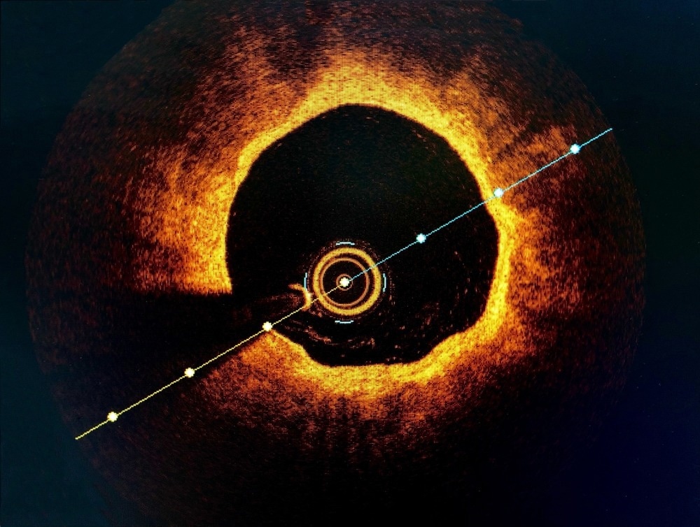
Image Credit: BelezaPoy/Shutterstock.com
Operating Principle
Optical coherence tomography enables the visualization of internal structures to capture detailed cross-sectional images of biological tissues. Optical coherence tomography operates on the principles of interferometry and low-coherence light, which involves directing a near-infrared light beam toward the target tissue.
A beam splitter divides this light into two paths: one directed towards a reference mirror and the other towards the biological tissue. The reflected light from both paths is then recombined, creating an interference pattern. By measuring the variations in the interference pattern, optical coherence tomography can construct detailed images of the internal structures with micron-level resolution.
Optical coherence tomography's main benefits are its high resolution, real-time image-capturing capability, and non-invasive design. Optical coherence tomography, in contrast to conventional imaging techniques like MRIs and X-rays, does not entail ionizing radiation and produces remarkably detailed cross-sectional images. This makes it especially useful for studying delicate structures like the retina, where accuracy is essential for diagnosis and treatment planning.
Applications of Optical Coherence Tomography
Optical coherence tomography finds diverse applications, especially in medical and biological fields. In ophthalmic imaging, OCT excels by non-contact delivery of light for high-resolution, 3-dimensional visualization of retinal and choroidal layers, surpassing traditional techniques. Optical coherence tomography angiography aids in diagnosing age-related macular degeneration, diabetic retinopathy, and glaucoma, crucial for sight-saving therapies.
In intravascular imaging, optical coherence tomography significantly enhances coronary artery disease diagnosis, guiding percutaneous coronary interventions with superior resolution and faster image acquisition compared to intravascular ultrasound. The optical biopsy provided by optical coherence tomography aids in the diagnosis and treatment planning of diseases such as Barrett's esophagus and colorectal polyps during gastrointestinal endoscopy.
Additionally, optical coherence tomography plays a pivotal role in pulmonary bronchoscopy, accurately assessing airway remodeling in obstructive lung diseases and aiding in the early diagnosis of lung cancer and interstitial lung disease.
Recent Studies
Enhancing Clinical Outcomes with OCT in Coronary Research
In a recent study on optical coherence tomography, significant progress has been made in understanding in vivo vascular biology. Over the past decade, optical coherence tomography has played a crucial role in identifying culprit plaque pathology, potentially revolutionizing the management of acute coronary syndromes.
The study emphasizes optical coherence tomography's capability to detect healed coronary plaque, contributing to insights into plaque destabilization and healing processes in atherosclerosis. Moreover, accurate detection of sequelae from percutaneous coronary interventions, often missed by angiography, could enhance clinical outcomes.
The study also underscores optical coherence tomography's essential role in diagnosing myocardial infarction with non-obstructive coronary arteries and its potential to improve understanding of neoatherosclerosis mechanisms, particularly in very late stent thrombosis cases.
Optimizing 3D Printing with FD-OCT
In a recent study, researchers introduced a novel application of Fourier-domain optical coherence tomography (FD-OCT) in multi-photon 3D laser printing. Traditionally, 3D microstructure inspection relied on ex-situ methods, causing slow optimization processes.
The study showcased an in-situ FD-OCT system integrated into a 3D laser lithography setup, allowing rapid assessment of 3D printed microstructures during the printing process. The FD-OCT system demonstrated the ability to examine polymer-monomer transitions and revealed time-dependent behavior after printing. The researchers emphasized the importance of non-invasive, in-situ monitoring techniques for identifying printing defects. This development enables fast, routine diagnostics without influencing the printing process and has the potential for optimizing 3D micro- and nanofabrication techniques.
Challenges and Future Prospects
Despite optical coherence tomography's success in certain medical fields, challenges like cost, limited field of view, and the need for dilated pupils still need improvements. The main challenge in optical coherence tomography lies in the interference-based artifact called speckle, which hinders consistent medical image interpretation.
Moreover, optical coherence tomography's limited depth of tissue penetration, especially in biological tissues, poses another challenge. Researchers are actively exploring solutions, including the development of advanced optical coherence tomography techniques and complementary imaging modalities to address these challenges.
Future prospects involve advancements in compact and affordable optical components, AI integration for automated diagnosis, and improving imaging speed and hardware integration for broader clinical use, especially in cardiovascular and gastrointestinal applications.
Conclusion
In conclusion, optical coherence tomography presents a transformative approach in medical imaging, offering non-invasive, high-resolution visualization of internal structures. It is used in ophthalmology, intravascular imaging, gastrointestinal endoscopy, and pulmonary bronchoscopy, improving diagnosis in various medical areas.
Recent studies highlight optical coherence tomography's pivotal role in understanding vascular biology and its potential in coronary atherosclerosis research. Another groundbreaking application in 3D laser printing shows the versatility of optical coherence tomography. The future of OCT looks bright with ongoing efforts to address the challenges, aiming for compact, affordable components, AI integration, and improved imaging speed for widespread clinical application.
More from AZoOptics: New X-Ray Technique Allows Effective Imaging of Living Organisms
References and Further Reading
Araki, M., Park, S. J., Dauerman, H. L., Uemura, S., Kim, J. S., Di Mario, C., ... & Jang, I. K. (2022). Optical coherence tomography in coronary atherosclerosis assessment and intervention. Nature Reviews Cardiology. https://doi.org/10.1038%2Fs41569-022-00687-9
Bouma, B. E., de Boer, J. F., Huang, D., Jang, I. K., Yonetsu, T., Leggett, C. L., ... & Wojtkowski, M. (2022). Optical coherence tomography. Nature Reviews Methods Primers. https://doi.org/10.1038%2Fs43586-022-00162-2
Mckenzie, Samuel. (2018, October 10). What is Optical Coherence Tomography?. News-Medical. Retrieved on December 13, 2023 from https://www.news-medical.net/health/What-is-Optical-Coherence-Tomography.aspx
Podoleanu, A. G. (2012). Optical coherence tomography. Journal of microscopy. https://doi.org/10.1111/j.1365-2818.2012.03619.x
Zvagelsky, R., Mayer, F., Beutel, D., Rockstuhl, C., Gomard, G., & Wegener, M. (2022). Towards in-situ diagnostics of multi-photon 3D laser printing using optical coherence tomography. Light: Advanced Manufacturing. https://doi.org/10.37188/lam.2022.039
Disclaimer: The views expressed here are those of the author expressed in their private capacity and do not necessarily represent the views of AZoM.com Limited T/A AZoNetwork the owner and operator of this website. This disclaimer forms part of the Terms and conditions of use of this website.