A team of researchers at the Massachusetts Institute of Technology (MIT) have designed a high-speed, three-dimensional imaging system to observe the pre-cancerous modifications in the colon or the esophagus.
The imaging system is based on the optical coherence tomography (OCT), a technology, which enables researchers to see microscopic details below the surface in three-dimension. The research has been published in the journal, “Biomedical Optics Express”.
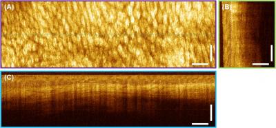 This is a 3-D OCT volumetric data set from an excised human colon specimen. (A) En face view showing regular organization of normal colon. (B and C) Cross-sectional views along two different directions showing sub-surface features. Two cross-sections are shown as examples, however multiple cross-sectional views can be extracted from the 3-D OCT data. Scale bar: 500um. Credit MIT
The imaging system developed by Professor James G Fujimoto and his team of researchers at MIT can capture data at 980 fps. Due to the fine resolution provided by the microscopic imaging system, the colon and esophageal cancer can be more easily diagnosed. A study by the American Cancer Society has determined that annually cancer affects over 1.5 million people each year.
In optical coherence tomography, light waves are used to examine the structures, which measure millionths of a meter in real-time. The technology has been applied in ophthalmology wherein by generating images of the retinal tissue, diseases such as glaucoma have been diagnosed and monitored.
Professor Fujimoto, who is also the senior author of the research paper titled, ‘Piezoelectric Transducer Based Miniature Catheter for Ultrahigh Speed Endoscopic Optical Coherence Tomography”, states that the imaging is of ultrahigh speed because it is capable of recording three-dimensional volumetric data of micron-scale resolution.
During the imaging process, pathological and structural features of a microscopic-scale are determined by directing a light beam over the tissue and calculating the echo time delay and magnitude of the backscattered light. For endoscopic OCT, optical probes that are a few millimeters wide are used to scan an optical beam and generate three-dimensional data sets of high resolution.
The team at MIT is collaborating with clinicians at the Harvard Medical School and the VA Boston Healthcare System to determine if the endoscopic OCT device can be used to guide excisional biopsy.