A novel technique has been developed for treating middle ear cholesteatoma using high-precision microsurgical ablation. This 445 nm semiconductor blue laser has been shown in ex vivo experiments to selectively remove diseased tissue to depths of up to 350 μm per pulse, while effectively preserving the underlying ossicular chain. These findings were published in Scientific Reports.
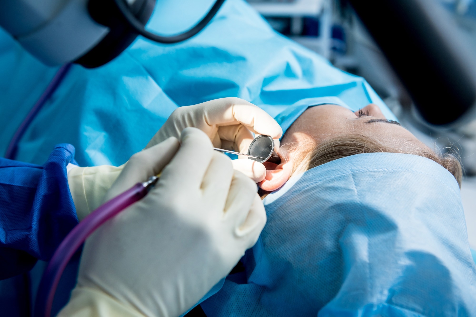
Study: Precise ablation of cholesteatoma using a 445-nm diode laser. Image credit: Roman Zaiets/Shutterstock.com
The findings address a long-standing clinical challenge by enabling targeted tissue removal within the middle ear, without causing mechanical damage or hearing loss in laboratory models.
The Clinical Challenge of Chronic Otitis Media
Chronic otitis media affects more than 20 million people worldwide, with approximately 25% of cases progressing to cholesteatoma, characterized by the growth of keratinizing squamous epithelium within the middle ear.
Standard treatment involves mastoidectomy procedures using mechanical drills and microsurgical instruments. However, because of the complex anatomy of the middle ear, these interventions can cause complications such as facial nerve injury, vertigo, and sensorineural hearing loss.
Limitations of Current Surgical Laser Technologies
To reduce these risks, various surgical lasers have been introduced. Infrared lasers, including the carbon dioxide (CO2) laser and Er:YAG laser, provide efficient tissue ablation due to their strong absorption by water. The cost and rigidity of specialized optical fibers limit their use in endoscopic microsurgery.
Visible wavelength systems, such as the KTP (potassium titanyl phosphate) laser, offer flexibility but have low hemoglobin absorption coefficients, necessitating higher power levels and increasing the risk of thermal damage to surrounding structures.
445 nm Diode Laser for Precise Ablation
To address the limitations of existing surgical laser systems, researchers investigated a semiconductor diode laser operating at 445 nm. This wavelength coincides with a strong hemoglobin absorption region, enabling efficient tissue ablation with shallow penetration depths and effective hemostasis.
The experimental setup employed a Fox IV diode laser with a 300 μm core diameter configured in single-pulse mode, delivering energy via a flexible multimode optical fiber.
The study evaluated laser-tissue interactions using three biological models: porcine ear cartilage as a surrogate for diseased ossicles, ex vivo human cholesteatoma tissue, and isolated human auditory ossicles, including the malleus and incus.
To simulate blood-rich conditions encountered during surgery, a controlled layer of diluted porcine blood was applied to all samples except the human-derived ossicles. This enabled blood-covered and non-covered tissue to be directly compared.
A 1060 nm spectral domain optical coherence tomography (SD OCT) system quantified ablation depths and crater morphology, providing lateral and axial resolutions of approximately 8 μm. Histological examination using hematoxylin and eosin (H&E) staining assessed tissue integrity and identified any thermal damage.
Click here to download a free PDF copy of this page
Impact of Laser Parameters on Tissue Ablation
The experiments showed a strong relationship between laser power and soft tissue ablation depth. Single pulses lasting 100 ms produced ablation depths of about 82 μm in human cholesteatoma tissue and 145 μm in the porcine model at low power settings of 0.5–1 W.
Increasing the power to 4 W raised the ablation depth to approximately 351 μm in human tissue and 372 μm in the animal model. Histological analysis confirmed that the underlying cartilage remained free of detectable structural or thermal damage across all soft tissue experiments.
The laser's effectiveness was influenced by fiber positioning. Increasing the distance between the fiber and tissue from 0.5 mm to 2 mm reduced energy density due to beam divergence, thereby decreasing ablation depth from approximately 260 μm to 170 μm at 2 W.
Changing the angle of incidence from shallow to perpendicular increased energy concentration and enhanced ablation efficiency. At 4 W, ablation depth increased from roughly 270 μm at 45 ° to a maximum of 435 μm at 90°.
A key finding was the laser's selective action on soft tissue relative to bone. At 1 W, the 445 nm laser effectively ablated cholesteatoma tissue while producing no detectable damage to human auditory ossicles.
Even at 4 W, bone ablation depths remained only 40–50% of those observed in soft tissue. This selectivity provides a substantial safety margin for preserving delicate middle ear structures, including the stapes crus, which typically measures only 0.3–0.8 mm in thickness.
Overlapping laser pulses with 26–42% spatial overlap produced continuous and uniform resection tracks with depths ranging from approximately 260 μm to 370 μm, without increasing peripheral thermal damage.
Implications for Endoscopic and Otological Surgery
The characteristics of the 445 nm diode laser make it a promising tool for microsurgical applications, particularly in middle ear surgery. Its compatibility with flexible optical fibers enables access to anatomically complex regions that are difficult to reach with conventional instruments. This capability may improve the removal of residual cholesteatoma tissue in confined areas of the middle ear.
The strong absorption of 445 nm light by hemoglobin also enables effective photoangiolytic hemostasis, thereby promoting rapid coagulation of small blood vessels and improving surgical visibility. Additionally, the blue wavelength may contribute to local microbial reduction, offering potential benefits in chronically infected middle ear environments.
Future Directions in Optical Theranostics
The study demonstrates that single-pulse 445 nm blue diode lasers can selectively ablate cholesteatoma tissue while preserving the delicate structures of the middle ear in ex vivo models.
By matching laser pulse duration to the thermal relaxation characteristics of the target tissue, thermal diffusion is confined to superficial regions, minimizing collateral damage and maintaining structural integrity.
The findings highlight a promising direction for future image-guided microsurgery. Integrating the 445 nm therapeutic laser with a 1060 nm OCT imaging system within a single endoscopic platform could enable real-time visualization of tissue morphology during treatment.
Such an approach would allow continuous monitoring of residual tissue thickness and dynamic adjustment of laser parameters, improving surgical precision and enhancing safety.
Journal Reference
Enzian, P., et al. (2026). Precise ablation of cholesteatoma using a 445-nm diode laser. Sci Rep 16, 15995. DOI: 10.1038/s41598-026-47908-6, https://www.nature.com/articles/s41598-026-47908-6
Disclaimer: The views expressed here are those of the author expressed in their private capacity and do not necessarily represent the views of AZoM.com Limited T/A AZoNetwork the owner and operator of this website. This disclaimer forms part of the Terms and conditions of use of this website.