*Important notice: This news reports on an unedited version of the paper which has been accepted and is awaiting final editing. Therefore, the study should not be regarded as conclusive or treated as established information.
Accurately reconstructing the complex wiring of the mammalian brain remains one of the biggest challenges in neuroscience. A recent study published in Scientific Data introduced an open-access dataset containing over eight million difficulty-graded annotation units derived from 10,547 neurons across 258 whole-brain mouse samples.
This repository is designed to train and evaluate artificial intelligence (AI) models for automated neuronal tracing, addressing a gap in standardized datasets necessary for effective algorithm development.
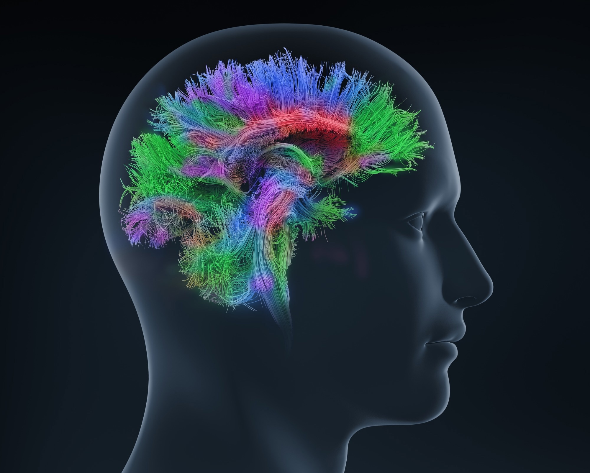 Study: A Million-Scale, Difficulty-Stratified Optical-Microscopy Image Annotation for Neuron Reconstruction. Image Credit: adike/Shutterstock.com
Enhancing Neuronal Reconstruction Techniques
Mapping complete axonal and dendritic projections at single-neuron resolution is key for understanding the functional architecture of the nervous system. Fluorescence micro-optical sectioning tomography (fMOST) enables high-resolution, three-dimensional (3D) imaging of entire brains while preserving the continuity of long-range neuronal projections. However, the large image datasets generated by whole-brain scanning create a processing bottleneck.
Saving this for later? Download a free PDF now.
Reconstructing neurons from petabyte-scale image stacks still relies heavily on manual tracing, which is slow and prone to variability. Automated tracing algorithms and deep-learning models have potential but are limited by the lack of large, standardized datasets.
Integrated Pipeline for Dataset Construction
To build this large-scale dataset, researchers developed an integrated pipeline combining high-resolution optical imaging, automated pre-tracing, and human verification. Neurons were first sparsely labeled using adeno-associated viral vectors to reduce signal overlap. The mouse brains were chemically stabilized, resin-embedded, and imaged with fMOST.
To maintain consistent image quality across the large volumetric scans, flat-field correction was applied to remove artifacts, and linear blending was used to eliminate stitching seams. The whole-brain images were divided into uniform blocks measuring 256 × 256 × 256 voxels, making them suitable for cloud-based processing.
Each block was pre-traced from the neuronal soma and evaluated using 11 quantitative metrics, including fiber density and structural complexity. These measurements were compressed using principal component analysis to calculate a difficulty score for each block, which was assigned to one of four difficulty levels.
The annotation workflow combined automated reconstruction with adaptive human verification. Junior annotators reviewed simpler blocks, while complex regions were assigned to experienced experts. Each block underwent independent checks by two annotators, with disagreements triggering a review process until consensus was achieved.
Comprehensive Dataset Overview
The completed HiNeuron V2 dataset contains 8,092,547 standardized annotation units from 10,547 neurons across 258 whole-brain mouse samples. It includes 201 samples acquired with high-definition fMOST and 57 collected using time-delay integration systems. Using principal component analysis, researchers then precisely grouped every image block into four difficulty levels, creating a benchmark for automated neuronal reconstruction.
Spatial analysis showed that challenging image blocks were concentrated around the neuronal soma, where branching is most complex, while simpler regions were more uniformly distributed throughout the neuronal arbor. Both axons and dendrites displayed similar difficulty, with level-one blocks accounting for about 66.3% of the dataset in axonal judgment data.
To evaluate the dataset, the study trained a 3D convolutional neural network (CNN) model using progressively larger training sets of 100, 400, and 1000 annotated image patches. Performance improved consistently as more training data was added.
For example, the tracing accuracy for level-one image blocks increased from 0.7553 to 0.8039 when the training set expanded from 100 to 1000 patches. This demonstrates that large, high-quality annotated datasets substantially enhance automated neuronal tracing.
Advancing AI-Driven Neuronal Tracing
This open-access dataset provides a valuable resource for developing, benchmarking, and improving automated neuron tracing algorithms. By pairing raw 3D optical microscopy images with verified neuronal skeletons, this approach provides a high-quality dataset for training and evaluating machine learning models. The difficulty-based organization helps researchers identify weaknesses in algorithms, enabling targeted improvements.
To support large-scale research, the repository includes interfaces for JavaScript, Python, and, Java, allowing users to filter and download data based on difficulty and sample size. This accessibility facilitates the evaluation of new algorithms for neuroscience and computer vision, thereby accelerating progress toward large-scale vertebrate connectome mapping.
Implications for Connectomics Research
The HiNeuron V2 dataset provides a standardized foundation for automated neuron tracing and connectomics research. By combining high-resolution fMOST with a difficulty-based annotation framework and collaborative human verification, researchers have addressed challenges related to data scale and annotation consistency.
As AI models continue to learn from this benchmark, the time needed to reconstruct complete mammalian projectomes could decrease. This scalable framework will enable faster, more accurate mapping of brain-wide neural circuits, helping scientists better understand both normal brain organization and the structural changes associated with neural circuit organization.
Journal References
Liao, M., et al. (2026). A Million-Scale, Difficulty-Stratified Optical-Microscopy Image Annotation for Neuron Reconstruction. Scientific Data. https://www.nature.com/articles/s41597-026-07668-4
Disclaimer: The views expressed here are those of the author expressed in their private capacity and do not necessarily represent the views of AZoM.com Limited T/A AZoNetwork the owner and operator of this website. This disclaimer forms part of the Terms and conditions of use of this website.