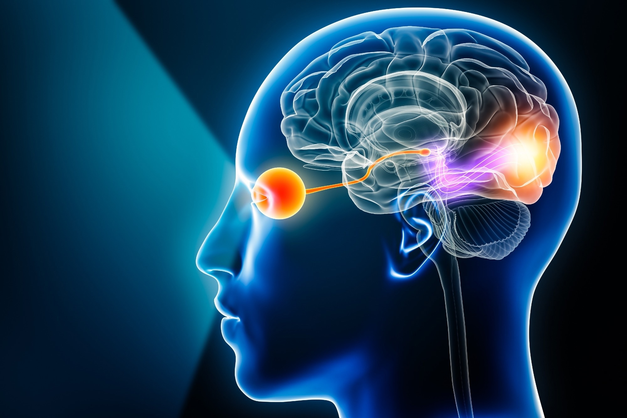
Animals and humans rely on an “internal compass” to navigate their environment. This compass is encoded by head direction (HD) cells, neurons that fire when the head points in a specific orientation. A new study from The Neuro (Montreal Neurological Institute-Hospital, McGill University) reports that visual objects sharpen the brain’s head-direction (HD) code by enhancing neurons tuned to the viewed direction and suppressing neurons tuned to other directions. This finding clarifies how vision stabilizes the brain’s internal compass and provides new insight into disorders where orientation is impaired.1

Image Credit: MattL_Images/Shutterstock.com
Background & Context
The internal compass that guides our sense of direction is rooted in a specialized network of neurons known as HD cells. These cells fire specifically when the head is oriented in a particular direction, or azimuth, regardless of the individual’s location.2,3 Working in concert with place cells in the hippocampus and grid cells in the entorhinal cortex, HD cells form a critical part of the brain’s navigation system, supporting the cognitive map that allows us to orient ourselves and move through space effectively.2
Earlier studies showed that HD cells can maintain tuning in the dark using idiothetic (vestibular and proprioceptive) inputs, suggesting that vision mainly calibrates an internally generated signal.3 However, landmark manipulations revealed that some HD populations reorganize in response to visual input, showing a more flexible role than originally assumed.4
Although visual cues were known to re-anchor HD activity after drift, it remained unclear whether objects actively refine the HD code or only reset orientation. The Neuro’s study addresses this by combining brain-wide mapping with targeted recordings to test how object viewing changes HD tuning.1,4
Accurate spatial orientation is essential for survival in animals and is one of the earliest cognitive abilities to decline in Alzheimer’s disease. Understanding how landmarks help stabilize the brain’s directional signal could offer valuable insight for early diagnostic tools.5 Beyond neuroscience, HD-like models inspired by these biological systems are also being applied in robotics and autonomous vehicles, where they integrate visual and inertial cues to support navigation in environments without GPS.6,7
Download the PDF of the article
Key Findings
The study reports three principal observations:
- Objects enhance aligned HD neurons. In the postsubiculum, a cortical HD hub, neurons tuned to the direction of a viewed object increased their firing, while neurons preferring other directions were suppressed. This selective modulation sharpened the population code for heading.1
- Object sensitivity is concentrated in navigation circuits. Brain-wide imaging showed that object-responsive signals were strongest in navigation areas, not in primary visual cortex. This suggests that object-based refinement is implemented where direction is computed.1
- Landmark properties matter. These findings are consistent with earlier work showing that the type and placement of landmarks (for example, symmetric versus asymmetric cues) can differentially anchor HD firing, with some HD subtypes more plastic than others.4,8
Overall, the results indicate that visual input does not simply provide calibration but actively tunes the neural compass to improve directional precision.1
Methodology
The researchers presented mice with controlled visual stimuli consisting of distinct objects or scrambled images that lacked object structure. Using whole-brain imaging, they identified a small number of regions where activity increased specifically when mice looked at objects. Among these, the postsubiculum showed the strongest responses, prompting targeted electrophysiological recordings. When objects were positioned at particular orientations, HD neurons tuned to those orientations increased their firing rates, while cells encoding other directions were inhibited. This combined approach, linking population-level mapping to single-cell analysis, demonstrated that objects refine HD coding within navigation circuits, rather than only re-establishing orientation after drift.1
Implications & Applications
In neuroscience and medicine, the findings highlight how impoverished or inconsistent landmarks can destabilize spatial orientation, a pattern also seen in the early stages of Alzheimer’s disease. This suggests that providing reliable landmarks may help support orientation in clinical environments, potentially offering a simple yet effective aid for patients experiencing early disorientation.5
In robotics, neural compass models benefit from object-based calibration to counteract drift caused by path integration. This approach enhances navigation accuracy in cluttered or GPS-denied environments, making systems more resilient and adaptable.6,7
For AR/VR systems, incorporating stable visual anchors in virtual spaces can help reduce spatial disorientation, improving overall user orientation and comfort during immersive experiences.
In the field of theoretical neuroscience, the results refine attractor-network models by showing that object inputs sharpen the tuning of head-direction cells within navigation circuits. This aligns with emerging evidence that not all HD cells follow perfectly coherent attractor dynamics, offering a more nuanced view of how these neural systems operate.4
Future Directions & Conclusion
Future research will need to explore whether similar mechanisms are at play in humans, how the complexity of landmarks affects orientation, and whether structured visual cues can be used therapeutically. From an engineering perspective, integrating object-aware visual modules into head direction (HD)-based navigation algorithms could boost reliability. Another key direction will be investigating multisensory integration, examining how auditory, tactile, or olfactory signals interact with visual objects to stabilize HD coding.
Long-term studies could also shed light on how consistent exposure to stable landmarks supports the consolidation of spatial maps over days or weeks. Extending this work to clinical populations may reveal whether thoughtfully designed environments can help reduce disorientation, offering potential benefits for individuals with dementia or vestibular disorders.
This study underscores how vision, and particularly the presence of objects, sharpens the brain’s internal compass by enhancing directional accuracy and anchoring internal estimates to the external world.1
Learn more about brain imaging here
References and Further Reading
- The Neuro (Montreal Neurological Institute-Hospital, McGill University). How visual cues fine-tune brain’s internal compass. EurekAlert! (10 September 2025). Available at: https://www.eurekalert.org/news-releases/1097560
- Taube, J.S. (2001). Neural Representations of Direction (Head Direction Cells). In Encyclopedia of Neuroscience. https://doi.org/10.1016/B978-0-08-097086-8.55037-5
- Sharp, P.E. (2010). Neural Representations of Direction (Head Direction Cells). In Encyclopedia of Neuroscience. https://doi.org/10.1016/B978-0-08-045396-5.00172-X
- Kornienko, O., Latuske, P., Köhler, L., Allen, K. (2018). Head-direction cells escaping attractor dynamics in the parahippocampal region. bioRxiv. https://doi.org/10.1101/268110
- Grieves, R.M., Shinder, M.E., Rosow, L., Leonard, A.S., Dudchenko, P.A. (2022). The Neural Correlates of Spatial Disorientation in Head Direction Cells. eNeuro, 9(6). https://doi.org/10.1523/ENEURO.0174-22.2022
- Li, B., Liu, Y., Lai, L. (2021). A Bio-Inspired 3-D Neural Compass Based on Head Direction Cells. IEEE Access, 9, 106–118. https://doi.org/10.1109/ACCESS.2021.3102477
- Chen, Y., Xiong, Z., Liu, J., He, Y., Li, M., Zhu, M. (2021). A Positioning Method Based on Place Cells and Head-Direction Cells for Inertial/Visual Brain-Inspired Navigation System. Sensors, 21(23), 7988. https://doi.org/10.3390/s21237988
- Sit, K.K., Goard, M.J. (2023). Coregistration of heading to visual cues in retrosplenial cortex. Nature Communications, 14, 1992. https://doi.org/10.1038/s41467-023-37704-5
Disclaimer: The views expressed here are those of the author expressed in their private capacity and do not necessarily represent the views of AZoM.com Limited T/A AZoNetwork the owner and operator of this website. This disclaimer forms part of the Terms and conditions of use of this website.