Optical spectroscopy techniques in proteomics extend beyond mass spectrometry by analyzing how proteins interact with light through absorption, emission, or scattering. These interactions report on sequence, structure, dynamics, and local environment in solution or on surfaces.
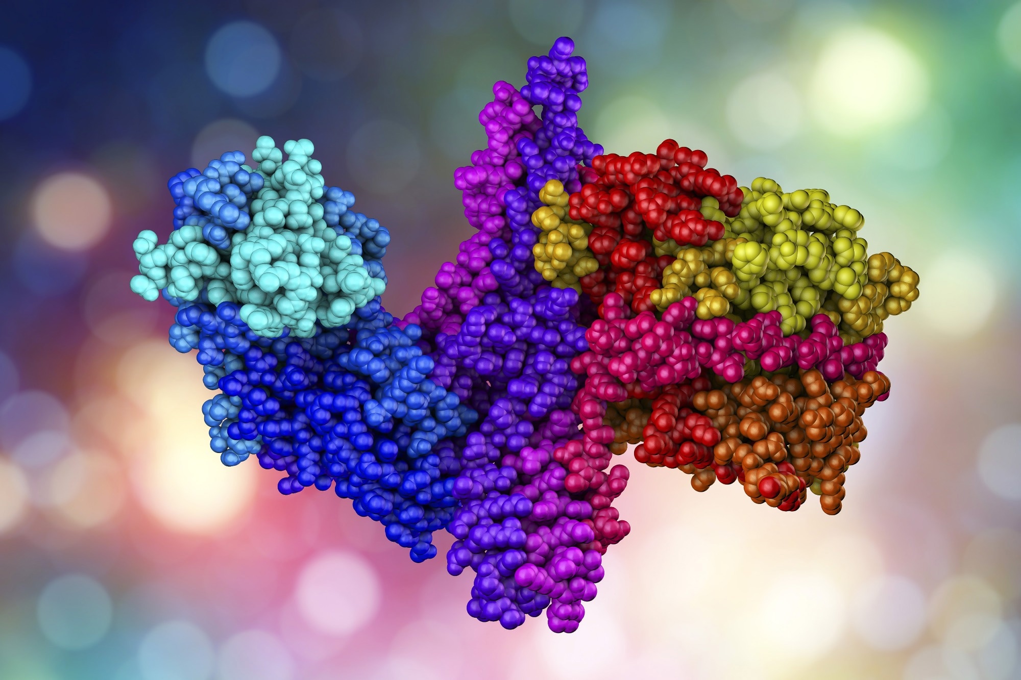 Image Credit: Kateryna Kon/Shutterstock
Optical methods complement mass spectrometry by capturing real-time behaviour under native conditions. Current research in protein analysis focuses on improving the extraction of information from optical signals through advanced detectors and single-molecule setups.
Additionally, the integration of microfluidics and nanopores enables direct sequencing, conformational analysis, and functional assays of intact proteins within complex environments.
Fluorescence Spectroscopy in Proteomics
Fluorescence spectroscopy is a powerful technique that measures light emitted by intrinsic protein residues like tryptophan or from extrinsic labels. Variations in intensity, lifetime, and anisotropy provide information about protein folding, binding events, and microenvironment polarity. Highly sensitive instruments enable precise quantitative comparisons of various protein states under controlled conditions.1,2
Labeling strategies attach dyes to specific amino acids or engineered tags, which creates position-resolved probes of large complexes. Single-molecule fluorescence methods increase the resolution of heterogeneity by monitoring individual proteins or complexes over time rather than relying on population averages.1,2
More advanced variants such as fluorescence correlation spectroscopy (FCS) resolve dynamics at the single-molecule level by analyzing intensity fluctuations in a tiny confocal volume. FCS yields critical data on diffusion coefficients, concentrations, and binding kinetics, making it invaluable for studies in membrane interaction proteomics and receptor-ligand interactions.3
Single Molecule Fluorescence and FRET
Single-molecule fluorescence techniques reduce the observation volume, allowing signals from individual protein molecules to be measured. This approach reveals rare states, intermediate conformations, and stochastic transitions that traditional ensemble methods average out. Such measurements are crucial for understanding molecular machines, folding pathways, and dynamic protein interactions.4
Förster resonance energy transfer (FRET) is another fluorescence-based technique that measures energy transfer between a donor and acceptor dye positioned at specific sites on a protein or protein pair. The efficiency of this energy transfer reveals distances at the nanometer scale. It allows the reconstruction of conformational changes, binding stoichiometry, and domain motions under varying biochemical conditions.5
Raman Spectroscopy and Surface-Enhanced Raman
Raman spectroscopy is an analytical technique that measures inelastic scattering of light. It directly probes vibrational modes of chemical bonds, yielding a label-free “molecular fingerprint” of proteins. Each band in the spectrum corresponds to specific backbone vibrations, side-chain modes, and secondary-structure elements so that Raman can distinguish between α-helix, β-sheet, and random-coil regions in purified proteins or complex mixtures.6
An advanced form of Raman spectroscopy, known as surface-enhanced Raman spectroscopy (SERS), amplifies signal strength by several orders of magnitude using metallic nanostructures. SERS can detect very low-abundance proteins or a small number of labeled molecules and allows ultra-sensitive, real-time, and multiplexed detection of proteins, biomarkers, and conformational changes.
Current developments focus on creating reproducible substrates, controlled surface functionalization, and integration with microfluidic systems for routine proteomic applications.6
Infrared and Two-Dimensional IR Spectroscopy
Infrared spectroscopy detects absorption of mid-infrared light that excites vibrational modes in the peptide backbone and side chains. The amide I and II bands are sensitive to secondary structure content such as alpha helices, beta sheets, and disordered segments. This sensitivity enables rapid assessment of global folding state and aggregation behavior in simple mixtures.7,8
Two-dimensional infrared spectroscopy introduces sequences of ultrafast pulses that correlate vibrational modes in frequency space. Cross peaks reveal couplings between structural elements and track how these couplings change over time. Such measurements provide detailed views of hydrogen bonding networks, folding kinetics, and the impact of ligand binding on protein energy landscapes.7,8
Circular Dichroism and Optical Activity
Circular dichroism (CD) spectroscopy measures the difference in absorption between left- and right-circularly polarized light by chiral molecules. Proteins exhibit distinct far-ultraviolet CD signatures that reflect their secondary structure composition. This property makes CD a valuable tool for quick quality control of refolded proteins, mutants, or formulated biotherapeutics.9
Near-ultraviolet CD probes tertiary structure by assessing the chiral environments of aromatic residues and disulfide bonds. Variations in these signals indicate local rearrangements that may not affect overall secondary structure fractions. Thus, CD complements infrared and Raman techniques in structural proteomics workflows, which track both global and local protein organization.9
Saving this for later? Download a PDF here.
Emerging proteomics workflows explore direct sequencing with optical readouts rather than mass analyzers. Some platforms rely on nanopores combined with fluorescent tags that report on specific amino acids as peptides thread through the pore. A study from Pacific Northwest National Laboratory describes concepts such as sequencing by degradation and reverse translation that convert sequence information into optical barcodes.10
These non-mass spectrometry platforms aim for high throughput, extended read capabilities, and straightforward instrumentation. Key challenges include managing peptide motion, developing reliable labeling chemistries, and constructing informatics pipelines that convert sparse optical patterns into precise sequences.
Recent studies demonstrate potential in specific applications but also point out limitations in depth compared to conventional liquid chromatography-mass spectrometry (LC-MS) workflows.10
Scattering and Elastic Light-Based Approaches
Elastic light scattering techniques, including single fiber elastic scattering spectroscopy, interrogate tissue and cell suspensions by measuring wavelength-dependent backscattered light. The resulting spectra reflect particle size, refractive index, and organelle density, which correlate with protein-rich structures and cytoskeletal organization.11
These scattering methods are well-suited for rapid, in situ assessment of protein-containing tissues, such as distinguishing benign from malignant regions based on shifts in scattering profiles. Because they do not require exogenous labels and can be implemented with fiber-optic probes, they are attractive for clinical and intraoperative proteomic monitoring where minimal sample preparation is critical.11
Integration with Mass Spectrometry and Future Directions
Optical spectroscopy techniques in proteomics often complement mass spectrometry rather than operating as isolated tools. Fluorescence or CD provides rapid feedback on sample quality, folding, or aggregation before digestion and mass spectroscopy analysis. Raman and infrared signatures classify samples or conditions, which guide targeted MS measurements for deeper coverage.10,12
Future work in optical spectroscopy aims for greater sensitivity, improved temporal resolution, and better integration with microfluidic handling and computational models. Meanwhile, non-mass-spectrometry sequencing concepts continue to evolve, and single-molecule optical studies refine biophysical models of complex proteoforms. Together, these trends expand what optical spectroscopy techniques in proteomics can reveal about protein function in native contexts.10
References and Further Reading
- Dos Santos Rodrigues, F. H. et al. (2023). Applications of fluorescence spectroscopy in protein conformational changes and intermolecular contacts. BBA Advances, 3, 100091. DOI:10.1016/j.bbadva.2023.100091. https://www.sciencedirect.com/science/article/pii/S2667160323000200
- Prasad, R. D. et al. (2025). A Review on Spectroscopic Techniques for Analysis of Nanomaterials and Biomaterials. ES Energy & Environment. DOI:10.30919/esee1264. https://www.espublisher.com/uploads/article_pdf/esee1264.pdf
- Melo, A.M. et al. (2014). Quantifying Lipid-Protein Interaction by Fluorescence Correlation Spectroscopy (FCS). In: Engelborghs, Y., Visser, A. (eds) Fluorescence Spectroscopy and Microscopy. Methods in Molecular Biology, vol 1076. DOI:10.1007/978-1-62703-649-8_26. https://link.springer.com/protocol/10.1007/978-1-62703-649-8_26
- Baraiya, K. A. et al. (2026). Chapter 14 - Fluorescence spectroscopy for single-molecule detection. Fluorescence Spectroscopy in Analytical Chemistry. DOI:10.1016/B978-0-443-30270-1.00025-5. https://www.sciencedirect.com/science/chapter/edited-volume/abs/pii/B9780443302701000255
- Verma, G. et al. (2025). Advances in FRET methodologies for probing molecular interactions. Biophysical Journal. DOI:10.1016/j.bpj.2025.08.027. https://www.cell.com/biophysj/fulltext/S0006-3495(25)00555-7
- Raman Spectroscopy in Protein Analysis. (2026). Nature. https://www.nature.com/nature-index/topics/l4/raman-spectroscopy-in-protein-analysis
- Hunt, N. T. (2024). Using 2D-IR Spectroscopy to Measure the Structure, Dynamics, and Intermolecular Interactions of Proteins in H2O. Accounts of Chemical Research, 57(5), 685. DOI:10.1021/acs.accounts.3c00682. https://pubs.acs.org/doi/10.1021/acs.accounts.3c00682
- Scherlo, M. et al. (2025). IR Spectroscopy: From Experimental Spectra to High-Resolution Structural Analysis by Integrating Simulations and Machine Learning. J. Phys. Chem., 129, 45, 11652–11665. DOI:10.1021/acs.jpcb.5c04866. https://pubs.acs.org/doi/10.1021/acs.jpcb.5c04866
- Oyama, T., Suzuki, S., & Akao, K. (2025). Circular dichroism spectroscopy in protein engineering and pharmaceutical development: Applications in structural characterization and quality assessment. Protein Expression and Purification, 237, 106826. DOI:10.1016/j.pep.2025.106826. https://www.sciencedirect.com/science/article/pii/S1046592825001688
- Deshpande, A. S. et al. (2025) Emerging protein sequencing technologies: proteomics without mass spectrometry? Expert Review of Proteomics, 22(3), pp. 89–106. DOI:10.1080/14789450.2025.2476979, https://www.tandfonline.com/doi/full/10.1080/14789450.2025.2476979
- Francis, F. et al. (2026). Advances in Light Scattering: A Comprehensive Analysis of Rayleigh, Dynamic, and Nonlinear Scattering Theories and Applications. Journal Of Advance And Future Research, Vol.4, Issue 1, page no.9-17. DOI:10.56975/jaafr.v4i1.502699. https://rjwave.org//jaafr/viewpaperforall.php?paper=JAAFR2601249
- Guo, T., Steen, J. A., & Mann, M. (2025). Mass-spectrometry-based proteomics: From single cells to clinical applications. Nature, 638(8052), 901-911. DOI:10.1038/s41586-025-08584-0. https://www.nature.com/articles/s41586-025-08584-0
Disclaimer: The views expressed here are those of the author expressed in their private capacity and do not necessarily represent the views of AZoM.com Limited T/A AZoNetwork the owner and operator of this website. This disclaimer forms part of the Terms and conditions of use of this website.