In a recent article published in the journal Biomedical Optics Express, researchers reported the design and evaluation of a refractive adaptive optics scanning light ophthalmoscope (AOSLO) specifically designed for small animal retinal imaging through a 1.8 mm pupil, with a focus on mice.
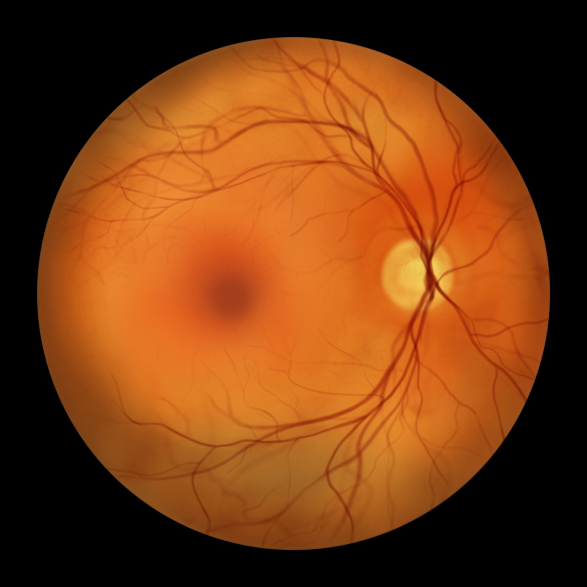
Image Credit: Kateryna Kon/Shutterstock.com
Why Small-Animal Retinal Imaging Needs a Rethink
Non-invasive optical imaging of the retina in animal models is essential for advancing understanding and treatment of various human eye diseases. Adaptive optics ophthalmoscopy traditionally relies on pairs of one-dimensional optical scanners arranged orthogonally to create raster scanning. This introduces complexity, lower light throughput, and wavefront aberrations.
Prior small animal AO systems have either used tilted spherical mirrors or refractive lens-based optics. While reflective setups avoid chromatic dispersion, they offer limited fields of view and present alignment challenges. Refractive systems allow broader retinal coverage but suffer from image degradation caused by lens reflections and require frequent optical alignment.
Additionally, existing scanners with resonant frequencies typically under 15 kHz limit temporal resolution. To address these limitations, this study explores a 2D MEMS scanner with a higher resonant frequency (~29.2 kHz) and minimal surface distortion, aiming to reduce system complexity, manage aberrations, and improve imaging speed.
Optical Design and System Architecture
The optical design used commercially available achromatic doublets selected from catalogues (Edmund Optics and Thorlabs) to form modified afocal relays that relay the pupil and retina conjugates with minimal aberrations. The final system comprised three such relays, imaging a 5.2 mm entrance pupil down to a 1.8 mm eye pupil.
The lens pairs were chosen based on low wavefront RMS error, diffraction-limited performance, and system length constraints (under 1 meter). Real ray tracing in Zemax simulated imaging quality, spot sizes, and aberrations across different focus settings and vergences without pre-compensation for species-specific aberrations such as spherical or longitudinal chromatic aberration.
To correct defocus and refractive errors, the team compared three approaches: wavefront correction using a pupil conjugate deformable mirror, a traditional Badal optometer adjusting object conjugates to the eye, and a modified Badal optometer that varies lens distances in the final relay. Each method's impact on image quality and wavefront errors was evaluated in a comparative framework.
The scanning system employed a 2D MEMS scanner with a 1.2 mm clear aperture and a high resonant frequency in one axis. This replaces the traditional pair of 1D scanners, reducing optical complexity and potentially improving light throughput.
The resonant axis operated near 29.2 kHz, while the orthogonal non-resonant axis operated at 50 Hz. To handle sinusoidal velocity variation and sampling jitter from the resonant scanning, three correction strategies were tested including direct analog orientation signal use, hybrid analog-digital schemes, and synthetic signal generation.
Imaging Performance and Optical Trade-Offs
Ray tracing simulations showed diffraction-limited imaging performance at 800 nm across the field of view and over a focus range sufficient to span the thickness of the mouse retina. Among the focus correction strategies, wavefront correction by the deformable mirror performed strongly under certain conditions, while the modified Badal optometer showed advantages in other regimes, supporting their complementary roles depending on refractive error.
The 2D MEMS scanner simplified parts of the optical layout by removing one pupil relay, increased throughput, and enabled higher frame rates compared to conventional 1D scanner configurations. However, operating the non-resonant axis at 50 Hz introduced deterministic and stochastic oscillations that limited the achievable resolution and stability.
Various correction schemes for scanner-induced image warping and line jitter were assessed. Hybrid and synthetic analog signals produced improved but not fully complete correction, while relying solely on noisy analog signals was insufficient for sub-pixel alignment.
Polarization methods effectively reduced lens reflections in imaging channels, with the combination of crossed polarizers and a quarter wave plate providing the strongest reflection suppression in the imaging path. This, however, introduced stronger corneal reflections that degraded wavefront sensor performance, necessitating additional illumination pattern adjustments.
Lens tilting also reduced reflections but at the cost of imaging aberrations and a reduced diffraction-limited focus range in traditional Badal configurations. The modified Badal correction helped recover this focus range despite magnification changes, highlighting a trade-off between reflection control and optical performance.
In vivo mouse retinal images demonstrated high resolution of multiple retinal layers using the refractive AOSLO system. Non-confocal quadrant detection provided complementary structural contrast to confocal images, revealing fine nerve fiber and vessel details.
Toward Faster, Simpler AO Retinal Imaging
This work presents a refractive adaptive optics scanning light ophthalmoscope optimized for small animal retinal imaging through a small pupil, employing commercially available achromatic doublets arranged in modified afocal relays. The study systematically evaluates optical strategies for lens reflection mitigation via polarization control and lens tilting, revealing practical trade-offs impacting both confocal imaging quality and wavefront sensing.
Overall, the refractive AO ophthalmoscope achieves diffraction-limited performance over relevant vergence and field ranges, enabling high-resolution, multi-modality in vivo imaging of mouse retina. The optical design principles, MEMS scanning implementation, and reflection control strategies provide a useful framework for future small-animal AO systems and related retinal imaging approaches, while retaining important system-level trade-offs.
Download the PDF of the article
Journal Reference
Karteek K., Yuning X., et al. (2026). Refractive adaptive optics scanning light ophthalmoscope with fast 2D MEMS scanner, Biomedical Optics Express 17, 635-655. DOI: 10.1364/BOE.576969, https://opg.optica.org/boe/fulltext.cfm?uri=boe-17-2-635