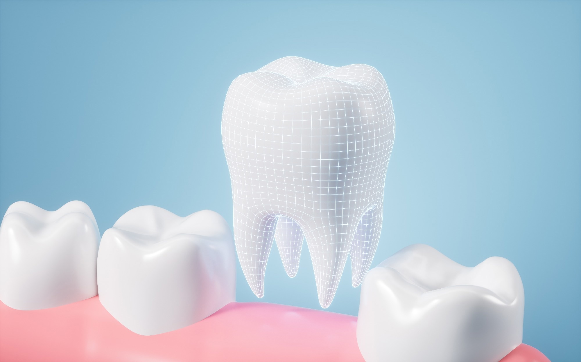
A recent article published in Scientific Reports explored the adhesion of primary human gingival fibroblasts and osteoblasts to orthodontic mini-implants (OMIs) using scanning electron microscopy (SEM).

Image Credit: Tina Ji/Shutterstock.com
The researchers aimed to understand how the surface microstructure of these implants affects cell adhesion and growth, which could lead to more effective and biocompatible orthodontic devices.
Background
OMIs are small titanium screws used in orthodontics to provide anchorage for tooth movement. They are usually placed in the jawbone and serve as temporary anchors. OMIs consist of three parts: the head (for attaching dental appliances), the neck (for close contact with the mucosa), and the threaded body (for fixation in the bone).
Unlike permanent dental implants, OMIs are designed to avoid tissue in-growth, making them easy to remove. They are primarily made from titanium alloys, known for their corrosion resistance and compatibility with bone.
OMIs often undergo surface treatments to enhance biocompatibility and cell adhesion. Treatments like ultraviolet (UV) light irradiation create a superhydrophilic surface that promotes cell attachment and proliferation. However, OMIs mainly rely on primary stability (macroretention) rather than secondary stability (osseointegration), ensuring easy removal.
About the Research
The authors investigated the adhesion of two different cell lines, primary gingival human fibroblasts and a human osteoblast cell line, to three commercially available OMIs: Tomas-pin, OrthoEasy Pin, and Dual Top G2 (primarily made from the titanium alloy TiAl6V4), which is widely used for orthopedic and dental implants.
They employed a combination of SEM and energy-dispersive X-ray (EDX) analysis to examine the surface microstructure and chemical composition of the implants. They then cultured the cells on the OMIs and observed their adhesion and growth patterns using SEM.
The study involved several steps. First, the OMIs were cleaned in 75 % ethanol using an ultrasonic bath and sterilized at 121 °C for 15 minutes. Next, EDX analysis was conducted to check the element composition of the OMIs. Then, SEM was used to examine the microstructure of the OMIs, focusing on the neck, thread, and head at 500× magnification.
Primary gingival fibroblasts and osteoblasts were then cultured on the OMIs, TC coverslips (positive control), and poly(1,1,2,2-tetrafluoroethylene) or PTFE (negative control) for comparison. Finally, SEM images at 500× magnification were taken to assess cell adhesion to the OMIs.
Research Findings
The SEM analysis showed varying surface microstructures, particularly in the neck and thread regions of the OMIs. The Tomas-pin and OrthoEasy Pin had smooth neck areas, while the Dual Top G2 had finely grooved structures. All threads had grooves, but the continuity and sharpness of the edges differed between the products.
Regarding cell adhesion, both fibroblasts and osteoblasts adhered to all areas of the OMIs, with cells aligning parallel to the grooves on the threads. Cell density was higher on grooved threads compared to smooth neck areas. The OrthoEasy Pin, despite its anodized oxide layer, showed no difference in cell adhesion compared to other OMIs, indicating that surface modification did not significantly affect cell response.
Similarly, the implant microstructure subtly influenced cell shape and spreading. Cells aligned parallel to the grooves, and higher cell density was observed on grooved surfaces, indicating a preference for microstructured surfaces.
Applications
The outcomes have implications for OMI design and optimization. Cell adhesion to the OMI head is undesirable as it can lead to connective tissue overgrowth, a common complication. However, fibroblast adhesion to the neck is beneficial, creating a tight seal around the implant and reducing infection risk.
Osteoblast adhesion to the thread region can enhance stability by promoting bone attachment, but excessive osseointegration could make removal difficult. Therefore, the current OMI surface microstructure achieves a good balance, supporting osteoblast adhesion without complete osseointegration.
Conclusion
In summary, the research provided valuable insights into how OMI surface microstructures affect the adhesion of gingival fibroblasts and osteoblasts. The results suggest that commercially available OMIs could influence cell behavior, with implications for their design and optimization.
While successful cell adhesion was observed, further surface modification could optimize cell behavior and minimize complications. Future work should focus on quantifying cell density and investigating the effects of different surface modifications on cell adhesion and osseointegration. These suggestions would help develop more effective, biocompatible OMIs, ultimately improving orthodontic treatment outcomes.
Journal Reference
Reimers, SNM., Es-Souni, M. Şen, S. (2024). Investigating adhesion of primary human gingival fibroblasts and osteoblasts to orthodontic mini-implants by scanning electron microscopy. Sci Rep. DOI: 10.1038/s41598-024-68486-5, https://www.nature.com/articles/s41598-024-68486-5
Disclaimer: The views expressed here are those of the author expressed in their private capacity and do not necessarily represent the views of AZoM.com Limited T/A AZoNetwork the owner and operator of this website. This disclaimer forms part of the Terms and conditions of use of this website.