Field-based scanning electron microscopy (SEM) is changing how surface imaging and microanalysis are conducted outside traditional laboratory environments.
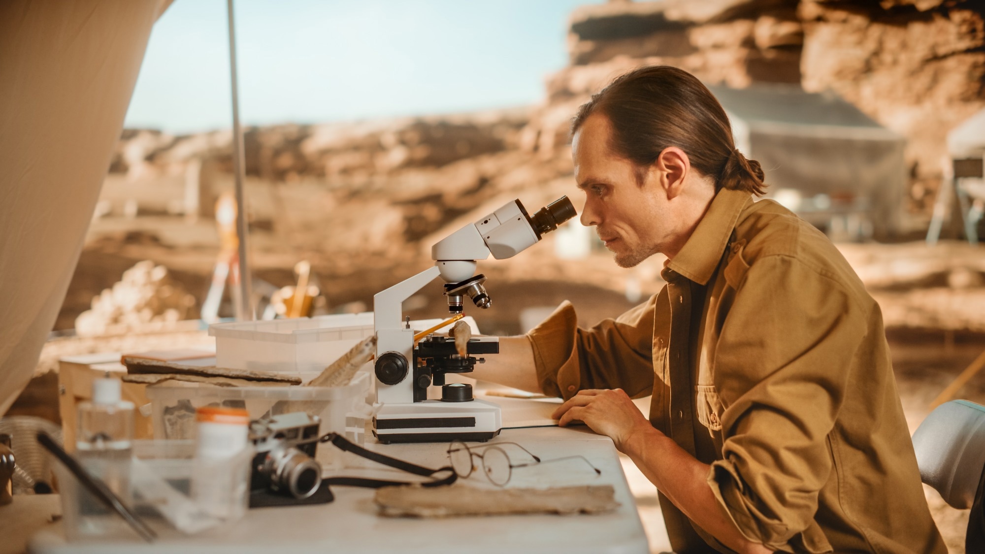
Image Credit: Gorodenkoff/Shutterstock.com
Unlike standard SEM systems, which are often large, stationary, and dependent on stable vacuum conditions, field-based SEMs are compact, mobile, and built for use in real-world environments.1
Across industries, there is a growing demand for non-destructive, in situ, and high-resolution imaging, particularly when delicate or time-sensitive samples can’t be easily transported. From archaeologists on remote digs to engineers investigating aircraft components on-site, portable SEM systems provide fast analysis where it's needed most.2
To meet these needs, field SEMs have been adapted with features such as smaller footprints for mobility, low-vacuum or environmental SEM (ESEM) modes for imaging uncoated samples, simplified software for ease of use, mobile power sources, and options for remote operation in hard-to-access areas.2,3
While these systems typically trade some resolution and sample size capacity for portability, their high utility in applied settings often outweighs those limitations. What they lack in pixel perfection, they make up for in accessibility, speed, and versatility.
Case Studies and Applications
Cultural Heritage Conservation
Field-based SEM is proving invaluable in cultural heritage conservation. It allows researchers to analyze historical pigments and artifact surfaces without removing samples from their original locations.
In one study, Falcon et al. used SEM combined with energy-dispersive X-ray spectroscopy (EDS) to map elemental compositions in natural soil pigments used in historic artworks.4
The team analyzed pigments such as Sienna yellow, burnt Sienna red, red earth, and green earth. SEM-EDS revealed unique elemental signatures—for example, burnt Sienna red had higher iron levels and lacked manganese, indicating a different firing process. Green and red earth pigments showed compositional differences linked to their geological origins.4
The study also included reference materials like GBW-07404 and NIST-SRM 2586, which showed silicate-dominant profiles with variable titanium and iron content. These distinctions offered insight into pigment provenance and helped inform both conservation strategies and authentication efforts.4,5
By enabling on-site, high-resolution analysis, field-based SEM minimizes sample handling and disruption, allowing conservators to preserve artifacts in situ while gaining critical data on their composition and history.
Download your PDF copy now!
Environmental Science: Tracking Microplastics with SEM
In environmental research, field-based SEM has become essential for studying microplastics (MPs)—small plastic fragments increasingly detected in marine ecosystems and food chains. Unlike standard imaging techniques, SEM provides high-resolution surface detail, helping researchers understand how environmental conditions affect plastic degradation over time.6
In a study by Miserli et al., MPs were extracted from seawater and the gastrointestinal tracts of fish. After identifying the polymer types using ATR-FTIR and micro-Raman spectroscopy, the team used SEM to examine surface morphology.
The micrographs showed that MPs—made from materials such as polyethylene (PE), polypropylene (PP), and polystyrene (PS)—had visibly rough, cracked surfaces with burrs and cavities.7
These irregularities contrasted sharply with the smooth surfaces of reference plastics, suggesting that environmental factors like mechanical wear, UV radiation, and microbial activity significantly alter plastic materials over time.7
Field-based SEM not only offers direct visual evidence of degradation but also helps distinguish different MP types, making it a practical, deployable tool for tracking plastic pollution and understanding its long-term ecological impact.
Mining and Geology
In mining and environmental remediation, field-based SEM plays a crucial role in real-time identification and characterization of minerals, particularly when assessing the success of in situ treatment strategies.8
One notable example involves the remediation of nickel-contaminated groundwater, where a fermentable carbon source was injected to promote microbially mediated precipitation of nickel sulfides (NiS).9
To track this process, Divine et al. used Min-Traps—sampling devices placed in monitoring wells for 64 days—to collect mineral precipitates formed during remediation. The retrieved samples were analyzed using a suite of techniques, including SEM-EDS. The resulting elemental maps clearly showed nickel and sulfur co-localized within the mineral structures.9
By enabling on-site mineral analysis, field-based SEM allowed researchers to confirm the geochemical transformation of contaminants and evaluate remediation performance without relying on delayed, off-site testing. More broadly, this example illustrates how portable SEM systems are enhancing field-based environmental monitoring and mineral exploration with timely, actionable insights.9
Forensics: Crime Scene Trace Evidence Analysis
Field-based SEM is gaining traction in forensic investigations for its ability to provide rapid, high-resolution analysis of trace evidence, particularly hair samples found at crime scenes. A frequent challenge in such cases is distinguishing human hair from animal hair, which can be critical in narrowing down suspects or validating testimony.
Traditional methods often relied on light microscopy to examine hair structure, but SEM offers much finer surface detail, making it possible to identify subtle morphological differences.10 For example, a study by O. F. Chernova used SEM to differentiate between human and primate hair with improved accuracy, offering a level of visual detail not achievable with conventional techniques.
While earlier forensic approaches focused on extracting nuclear DNA from hair shafts, success was limited due to the naturally low DNA content. Mitochondrial DNA (mtDNA), which is more abundant in hair, has since become a valuable tool in degraded or older samples.11
The key advantage of portable SEM systems in this context is their ability to deliver high-resolution imaging on-site, minimizing contamination risks and enabling faster analysis without needing to transport delicate samples to a lab. This is particularly useful at remote or time-sensitive crime scenes, where every hour can impact the investigation.10
Outlook and Opportunities
The use of field-based SEM is expanding quickly, driven by growing interest in mobile lab deployments, on-site diagnostics, and real-time analysis in remote settings. These portable systems are increasingly valuable in areas like disaster response, quality control at production sites, and autonomous environmental monitoring.2
As integration with cloud platforms and AI-powered image analysis advances, remote collaboration and automated reporting are becoming more accessible. These tools also hold strong potential in education and field training, enabling hands-on learning without the need for traditional lab infrastructure.1,2
Developments may focus on boosting resolution, extending battery life, enhancing wireless connectivity, and incorporating machine learning for automated pattern recognition. As hardware continues to shrink and software becomes more user-friendly, field-based SEMs are well-positioned to move from specialist use to standard practice across various sectors.
For more on in-field innovation and portable diagnostics, explore:
Curious about just how compact field-based SEMs can be?
The World's Smallest Scanning Electron Microscope
References and Further Reading
1. Zhou, W.; Greer, H. F., What Can Electron Microscopy Tell Us Beyond Crystal Structures? European Journal of Inorganic Chemistry 2016, 2016, 941-950. https://chemistry-europe.onlinelibrary.wiley.com/doi/full/10.1002/ejic.201501342
2. Ali, A.; Zhang, N.; Santos, R. M., Mineral Characterization Using Scanning Electron Microscopy (Sem): A Review of the Fundamentals, Advancements, and Research Directions. Applied Sciences 2023, 13, 12600. https://www.mdpi.com/2076-3417/13/23/12600
3. Lewczuk, B.; Szyryńska, N., Field-Emission Scanning Electron Microscope as a Tool for Large-Area and Large-Volume Ultrastructural Studies. Animals 2021, 11, 3390. https://www.mdpi.com/2076-2615/11/12/3390
4. Falcone, F.; Cinosi, A.; Siviero, G.; Rosatelli, G., Innovative Methodological Approach Integrating Sem-Eds and Txrf Microanalysis for Characterization in Materials Science: A Perspective from Cultural Heritage Studies. Spectrochimica Acta Part B: Atomic Spectroscopy 2024, 218, 106980 DOI: https://doi.org/10.1016/j.sab.2024.106980.
5. Bacon, J. R.; Butler, O. T.; Cairns, W. R.; Cook, J. M.; Mertz-Kraus, R.; Tyson, J. F., Atomic Spectrometry Update–a Review of Advances in Environmental Analysis. Journal of Analytical Atomic Spectrometry 2019, 34, 9-58. https://pubs.rsc.org/en/content/articlelanding/2022/ja/d1ja90054d
6. Thomas, C.; Spatayeva, T.; Yu, D.; Loh, A.; Yim, U. H.; Yoon, J.-Y., A Comparison of Current Analytical Methods for Detecting Particulate Matter and Micro/Nanoplastics. Applied Physics Reviews 2024, 11. https://doi.org/10.1063/5.0153106
7. Miserli, K.; Lykos, C.; Kalampounias, A. G.; Konstantinou, I., Screening of Microplastics in Aquaculture Systems (Fish, Mussel, and Water Samples) by Ftir, Scanning Electron Microscopy–Energy Dispersive Spectroscopy and Micro-Raman Spectroscopies. Applied Sciences 2023, 13, 9705. https://www.mdpi.com/2076-3417/13/17/9705
8. Tusa, L.; Frenzel, M.; Pereira, L.; Thiele, S.; Tolosana-Delgado, R.; Gutzmer, J., Geometallurgy: Future Directions. SEG Discovery 2025, 27-39 DOI: 10.5382/Geo-and-Mining-26. https://doi.org/10.5382/Geo-and-Mining-26
9. Divine, C.; Justicia-León, S.; Tilton, J. M.; Carter, E.; Zardouzian, E.; Clark, K.; Taggart, D., Field Methods and Example Applications for the Min-Trap® Mineral Sampler. Remediation Journal 2023, 33, 209-216 DOI: https://doi.org/10.1002/rem.21752.
10. Morillas, A. V.; Suhling, K.; Frascione, N., Unlocking the Potential of Forensic Traces: Analytical Approaches to Generate Investigative Leads. Science & Justice 2022, 62, 310-326. https://www.sciencedirect.com/science/article/pii/S1355030622000417
11. Chernova, O. F., Scanning Electron Microscopy of the Hair Medulla of Orangutan, Chimpanzee, and Man. Doklady Biological Sciences 2014, 456, 199-202 DOI: 10.1134/S0012496614030065.
Disclaimer: The views expressed here are those of the author expressed in their private capacity and do not necessarily represent the views of AZoM.com Limited T/A AZoNetwork the owner and operator of this website. This disclaimer forms part of the Terms and conditions of use of this website.