A recent study published in the journal Advanced Science introduced Lipi-PS, a wavelength-ultrasensitive fluorescent probe to enhance hyperspectral fluorescence imaging (HSFI). This novel probe enables high-resolution mapping of microenvironmental polarity and real-time tracking of lipid droplets (LDs) in live cells, tissues, and zebrafish through five-dimensional (5D) tracking, encompassing spatial (three-dimensional or 3D), spectral, and temporal dimensions.

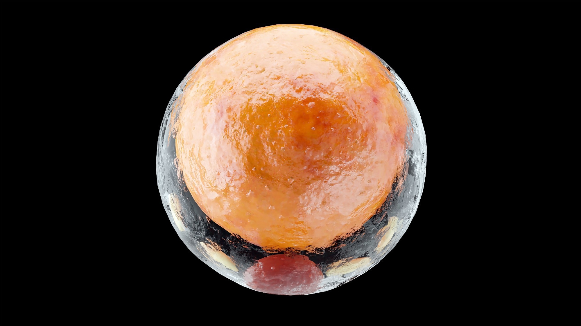
Image Credit: ALIOUI MA/Shutterstock.com
The finding highlights Lipi-PS as a new benchmark for optics-based bioimaging, offering unprecedented sensitivity for visualizing intracellular dynamics at the sub-organelle level.
Advancements in Fluorescence Imaging Technology
Fluorescence imaging is important in biomedical research due to its non-invasive nature and high sensitivity. Traditional methods, such as wide-field and confocal microscopy, provide structural information but often lack detail about the microenvironment.
HSFI captures the full fluorescence spectrum at each pixel, allowing for the analysis of microenvironmental changes based on spectral shifts. However, this method has been limited by the availability of highly wavelength-sensitive fluorescent probes. Common dyes such as Nile Red show very small spectral shifts (approximately 7 nm) between different solvents, making them unsuitable for detecting subtle changes in biological systems.
Developing a Probe for Enhanced Polarity Sensitivity
The study synthesized and evaluated 12 donor-π-acceptor (D-π-A) molecules by varying donor groups, π-spacers, and acceptors to overcome sensitivity limitations. Fluorescence spectroscopy and quantum calculations showed that the π-spacer plays a dominant role in enhancing polarity sensitivity. Among the candidates, compound 12, featuring a fluorene π-spacer, benzodithiophene tetra-oxide acceptor, and NEt2 donor, achieved an exceptional polarity slope (Kps) of 27.1, far surpassing conventional dyes.
Researchers developed Lipi-PS based on this scaffold, incorporating cyano-phenyl groups for red-to-near-infrared (NIR) emission and alkyl chains for selective lipophilic targeting. Lipi-PS demonstrated strong absorption at 550 nm and exceptional solvatochromic emission, shifting from 617 nm in nonpolar hexane to 888 nm in polar dichloromethane. This corresponds to a polarity slope of 29.8, about six times higher than Nile Red's 4.9, marking the highest sensitivity reported among polarity-sensitive fluorescent probes.
Computational orbital analysis showed significant charge separation, highlighting Lipi-PS's strong polarity sensitivity. As solvent polarity increases, the dihedral angle between the π-spacer and acceptor decreases, enhancing π-conjugation and shifting emission wavelengths. Additionally, Lipi-PS achieved a quantum yield of 38.6% in low-polarity solvents, which is remarkably high for dyes operating in the NIR window.
Demonstrated Optical Sensitivity and Dynamic Spectral Imaging
The Lipi-PS probe outperformed conventional LD stains like Nile Red in specificity, signal-to-noise ratio (SNR), and photostability. It achieved an SNR of 34.1 ± 13.9, compared to 2.6 ± 2.2 for Nile Red, and retained over 92% fluorescence intensity after 100 confocal scans.
Using wavelength-scanning confocal microscopy combined with MATLAB-based spectral decoding, researchers established a 4-step HSFI polarity mapping technique. Each pixel's emission spectrum was correlated to solvent polarity using the ET(30) scale, enabling quantitative mapping of subtle microenvironmental polarity changes.
In HeLa cells treated with polarity-altering agents, HSFI demonstrated that cholesterol reduced LD polarity, while oleic acid increased it. LD sizes shifted accordingly, reflecting compositional changes between cholesterol esters and triglycerides.
Lipi-PS also differentiated cancerous (HeLa, HepG2) and non-cancerous (HT22, 7702) cells based on LD polarity, highlighting its potential in tumor diagnostics. In mouse liver tissues, HSFI visualized LD polarity differences between healthy and non-alcoholic fatty liver disease models, showing polarity decreases and LD size increases during disease progression.
HSFI tracked polarity dynamics during zebrafish development at the whole-organism level, indicating organ-specific variations. A particular application involved 3D single-molecule fluorescence spectral dynamics imaging microscopy, enabling 5D tracking of LDs in live cells. This approach captured spatial positioning and spectral polarity changes, indicating dynamic compositional shifts and intracellular diffusion behaviors at the single-droplet level.
Optical Applications Across Cellular and Organismal Models
The robust spectral sensitivity of Lipi-PS enables precise subcellular polarity mapping by visualizing biochemical variations in lipid droplets. It distinguishes tumorigenic from non-tumorigenic tissues based on lipid composition and polarity, demonstrating strong potential for disease diagnostics. Lipi-PS supports 5D imaging, allowing real-time tracking of organelle movement and microenvironmental changes in cells and zebrafish. Its photostability, near-infrared emission, and low cytotoxicity make it ideal for long-term live-cell tissue imaging.
By overcoming the key limitation of wavelength sensitivity in HSFI, Lipi-PS serves as a benchmark for polarity-sensitive probes. Its ability to correlate spectral shifts with lipid droplet composition opens new directions for studying lipid metabolism and disease progression, such as non-alcoholic fatty liver disease (NAFLD) and developmental biology. The integration of HSFI with super-resolution techniques such as stimulated emission depletion (STED) microscopy could further enhance high-resolution biological imaging.
Conclusion and Future Directions for Optical Biosensing
Lipi-PS establishes a new benchmark for polarity-sensitive fluorescent probes, overcoming sensitivity limitations that previously restricted HSFI. Its combination of high quantum yield, long-wavelength emission, strong two-photon absorption, and robust photostability addresses key challenges in bioimaging, enabling precise real-time visualization of physiological and pathological processes at the subcellular level.
The successful application of Lipi-PS in mapping lipid droplet polarity across cells, tissues, and whole organisms highlights its versatility in biomedical research. This advancement enhances HSFI capabilities and opens new directions for investigating lipid metabolism and cellular microenvironment dynamics. The continued evolution of probe chemistry and the adoption of Lipi-PS will make HSFI a useful tool in biomedical optics and life sciences.
Disclaimer: The views expressed here are those of the author expressed in their private capacity and do not necessarily represent the views of AZoM.com Limited T/A AZoNetwork the owner and operator of this website. This disclaimer forms part of the Terms and conditions of use of this website.
Source:
Zhou, R., & et al. Hyperspectral Fluorescence Imaging with a New Polarity-Ultrasensitive Fluorescent Probe. Advanced Science, e08792 (2025). DOI: 10.1002/advs.202508792, https://advanced.onlinelibrary.wiley.com/doi/10.1002/advs.202508792