A diagram to show the basic overview of the system. Firstly, the sample is labeled with the photoswitchable Raman probe. It’s then irradiated with two-color infrared laser pulses, ultraviolet light, and a special donut-shaped beam of visible light to constrain the area where Raman scattering can occur. As a result, the probe can be detected at a very precise point for high spatial resolution images. Image Credit: 2023 Ozeki et al. CC-BY
In the microscopic world, humans have employed optical devices to peer in ever-increasing detail for as long as humanity has been able to manipulate light.
Modern microscopic imaging techniques go far beyond what conventional microscopes can provide. Two prominent technologies are super-resolution fluorescence imaging, which provides good spatial resolution, and vibrational imaging, which compromises spatial resolution but can utilize a wide array of colors to aid in labeling several types of constituents in cells.
We were motivated by the limitations of these kinds of imaging techniques to try and create something better, and with RESORT we are confident that we have achieved this. RESORT stands for reversible saturable optical Raman transitions, and it combines the benefits of super-resolution fluorescence and vibrational imaging without inheriting the detriments of either. It is a laser-based technique that uses something known as Raman scattering, a special interaction between molecules and light which helps identify what’s in a sample under the microscope. We successfully performed RESORT imaging of mitochondria in cells to validate the technique.
Professor Yasuyuki Ozeki, Research Center for Advanced Science and Technology, University of Tokyo
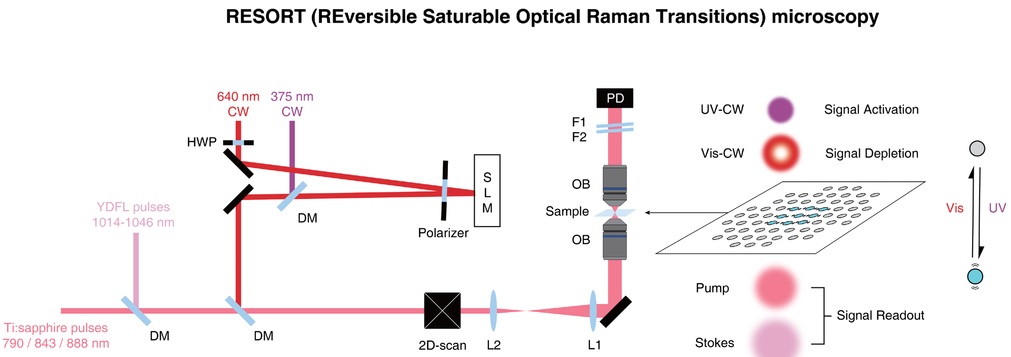
There are many stages to RESORT imaging, and even though it might seem complex, the setup is less complex compared to that of the techniques it is trying to exchange. At first, the particular components of the sample to be imaged have to be stained or labeled with special chemicals known as photoswitchable Raman probes, whose Raman scattering can be regulated by the varied types of laser light used by RESORT.
Next, the sample is kept within an optical apparatus that is employed to appropriately illuminate the sample and develop an image of it. For that to happen, the sample is then irradiated using two-color infrared laser pulses for detecting ultraviolet light, Raman scattering, and a special donut-shaped beam of visible light. Overall, these restrict the area where Raman scattering can happen, which implies the final stage, imaging, can diagnose the probe at a very accurate point, resulting in a high spatial resolution.
It’s not just about gaining higher-resolution images of microscopic samples; after all, electron microscopes can image these things in far greater detail. However, electron microscopes necessarily damage or impede the samples they observe. Through the future development adding more colors to the palette of Raman probes, RESORT will be able to image many components in living samples in action to analyze complex interactions like never before. This will contribute to a deeper understanding of fundamental biological processes, disease mechanisms, and potential therapeutic interventions.
Professor Yasuyuki Ozeki, Research Center for Advanced Science and Technology, University of Tokyo
The key focus of the team was to enhance microscopic imaging for application in the medical research field and related areas. However, the progress it has made in laser design could be utilized in other laser applications as well, where accurate control or high power is needed, such as materials science.
Journal Reference
Shou, J., et al. (2023) Super-resolution vibrational imaging based on photoswitchable Raman probe. Science Advances. doi.org/10.1126/sciadv.ade9118.