In a recent study published in Optica, scientists at Lawrence Livermore National Laboratory (LLNL) created a 3D quantum ghost imaging microscope, the first of its type.
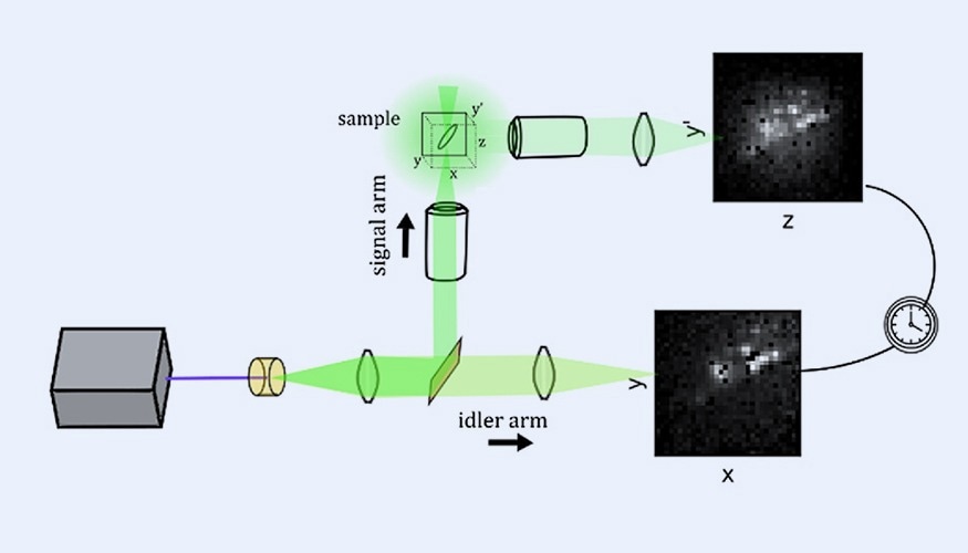 The 3D quantum ghost imaging microscope setup. A laser and crystal (left) are used to make entangled photons, which are split and sent in two directions. One turns left to hit and scatter off a sample, providing a standard image at a 90-degree angle. The other continues straight and is used to construct a ghost image. Image Credit: Eshun et al.
Until now, two dimensions or two planes at fixed z locations have been the only options for quantum ghost imaging.
This is a new way of 3D imaging that can do things with more sensitivity and gather more information without having to scan a sample.
Audrey Eshun, Study Author and Scientist, Lawrence Livermore National Laboratory (LLNL)
The strategy relies on the quantum phenomena of entanglement. A laser lights a crystal, resulting in photon pairs that are entangled or connected in space and time. These couples collide with a mirror that separates them: the "signal" photon moves left toward the sample, while the "idler" photon proceeds straight to a camera-like detector.
Idler photons that do not interact with the sample produce a homogeneous, featureless image on the detector.
Meanwhile, signal photons pass via a microscope objective, which gathers and focuses them on the sample. In this study, the scientists investigated metallic nanoparticle clusters.
The sample is angled 45 degrees relative to the incoming photons. When photons hit it, they scatter in all directions.
Another microscope objective, positioned at a right angle to the incoming light, gathers scattered photons and sends them to a second detector. This camera produces a typical picture of the nanoparticles' y-z plane.
Both detectors determine the precise arrival time of each photon. By comparing the timestamps of photon pairs received by both cameras, researchers may identify which idler photons correspond to signal photons that interact with the sample. They erase every other photon from the featureless idler picture, leaving just a ghost image of the sample's x-y plane.
“The standard image has y and z coordinates and a time for each pixel, and the ghost image has x and y coordinates and a time for each pixel. By grouping all the photons that have the same timestamp, we can figure out the x, y and z position for each photon. These coordinates can then be plotted to form a 3D image.” added Eshun.
In contrast to previous approaches, 3D quantum ghost imaging does not need to scan the sample and may occur all at once. It employs extremely low light intensities, making it suitable for imaging light-sensitive materials.
This microscope is the first of its kind. There was another 3D quantum ghost image, but in that case the resolution was about 3 cm. This is microns. We are getting three spatial dimensions of information at the micron scale.
Ted Laurence, Study Author and Scientist, Lawrence Livermore National Laboratory (LLNL)
The team's next goal is to apply this technology to the high-speed tracking of cell movements relative to one another.
The Biological and Environmental Research Program of the United States Department of Energy provided funding for this study.
Journal Reference:
Eshun, A., et al. (2025). 3D quantum ghost imaging microscope. Optica. doi.org/10.1364/OPTICA.565248.