Researchers have developed a system that uses dual-wavelength light to provide information about tumor depth in healthy tissue. Image Credit: Christine M. O’Brien, Washington University in St. Louis
During tumor removal, doctors can utilize fluorescent chemicals that make cancer cells glow so the surgeon can observe if any malignant tissue is still present. However, the equipment required for this method is not easily available and frequently does not offer quantitative data regarding how deeply the cancer cells are buried in the tissue.
Access to detailed information would aid surgeons in removing the entire layer of healthy tissue surrounding the tumor, which has been demonstrated to result in optimal patient outcomes.
The few commercial systems that do provide quantitative depth information are large and expensive, limiting use outside of large medical centers. Our group built upon prior work in this field to develop a low-cost, simple system that can quickly determine the depth of tumor cells using near-infrared (NIR) fluorescent probes.
Christine M. O’Brien, Research Team Leader and Instructor, Washington University School of Medicine in St. Louis
The journal Biomedical Optics Express from the Optica Publishing Group includes an overview of the researchers' new system. Utilizing the portable and user-friendly device in clinical settings with limited resources could reduce health inequities.
“Systems like this could be used in the future to improve surgical outcomes of patients undergoing tumor removal. It would also prevent the need to wait for pathology results before confirming whether cancer cells are still present after tumor removal,” O’Brien stated.
Lighting Up Cancer
According to studies, surgical cancer treatments are more likely to be successful if surgeons completely remove both the tumor and the healthy layer of tissue surrounding it.
However, this can be challenging because it is difficult to define the borders between the tumor and healthy tissue. Additionally, the type and location of the tumor affect the ideal thickness of the healthy layer.
The Achilefu Lab research team, under the direction of O’Brien, created a new tool to aid in this endeavor. It is based on applying a single fluorescent dye during tumor resection, which can subsequently be activated by two separate NIR-wavelengths that penetrate different depths in the tissue.
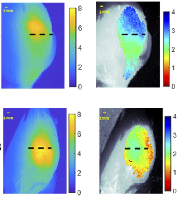
Cancer cells can be found 1 to 2 cm below the surface, thanks to the NIR-fluorescence that is released and can be viewed through tissue.
Dual wavelength excitation fluorescence makes use of the fact that different light colors or wavelengths go through tissue at various speeds. The depth to which tumor-targeted agents are positioned within the tissue can be determined by illuminating fluorescent molecules that target tumors with various light wavelengths and observing how they react.
O’Brien stated, “Multiple research groups have contributed to the development of mathematical relationships that link fluorophore depth to ratiometric fluorescence measurements. The surge of near-infrared contrast agents being developed for use in medicine encouraged us to build upon prior work and to create a system that works in the near-infrared and that is also low-cost and simple to use.”
Building a Dual-Wavelength System
The two wavelengths of excitation light used in the innovative fluorescence imaging system are 730 nanometer and 780 nanometer LEDs, and a monochrome CMOS camera is used to detect the ensuing fluorescence. To match the fluorescence images with the actual view of the tissue, a brightfield image was also made using an 850-nanometer LED.
Due to its broad excitation spectrum and capacity to be administered during tumor resection, the experimental drug LS301, developed in the Achilefu Lab, was chosen by the researchers as the cancer-targeting infrared probe.
This decision was made because using more than one fluorophore would have complicated the clinical application. Clinical trials with LS301 are now being conducted on people with breast cancer.
The system was examined using chicken slices and layered synthetic materials, and the researchers next evaluated its capacity to predict tumor depth using mouse breast tumors. The mice were first injected with LS301, and then the system was used to image them.
The required images were captured in five minutes. Calculations made using these images showed a good correlation with the true depth of the tumor and an average error of just 0.34 mm, which is probably acceptable for clinical application.
By accelerating data processing and adding more automation to the system so that it can scan the entire tissue surface, the researchers are currently striving to make the technology even more beneficial for surgical guidance.
Journal Reference:
O’Brien, C. M., et al. (2022) Quantitative tumor depth determination using dual wavelength excitation fluorescence. Biomedical Optics Express. doi:10.1364/BOE.468059.