Two-photon excitation microscopy (TPEF or 2PEF) is a crucial tool in biological imaging. By exciting fluorescent markers utilizing lower-energy photons and influencing nonlinearities of the absorption process, TPEF delivers numerous advantages, including reduced scattering, reduced photobleaching and increased imaging depth.
Fluence Technology developed Halite, a series of class-leading femtosecond lasers, to offer cost-efficient solutions for the most exacting of applications.
The Advantages of Two-Photon Excitation Microscopy
Since its inception in the 1990s, TPEF has become a powerful and extensively-used biological imaging technique.1 The foundation of all fluorescence microscopy methods is fluorescent markers (known as fluorophores), which can be customized to bind to particular sites and structures in intricate biological samples and live tissues, such as individual cells of a genetically defined type.
Moreover, fluorescent markers display structures of samples, high-contrast morphology and tissues imaged via two-photon microscopes by emitting visible light. Fluorophores must first be excited in order to emit visible light. Traditionally, a photon with higher energy (shorter wavelength) than the emitted light excites each chromophore in microscopy applications.
This method is subverted when using TPEF by using two relatively low energy (long wavelength) photons whose combined energy roughly matches the energy of the emitted photon to achieve the required excitation.
Excitation is initiated when these two photons are simultaneously absorbed by a fluorophore, leaving a molecule at a higher energy level. There are many benefits of exciting a fluorophore utilizing lower-energy photons.2
Since scattering increases with energy, two-photon techniques display reduced scattering and resultingly offer enhanced imaging depth and suitability for in-vivo imaging.3 Moreover, the photobleaching of the sample is lowered.
Also, another advantage offered by TPEF is thanks to the combination of the nonlinearity of the process and laser scanning mechanisms which delivers higher resolution in thicker samples than conventional fluorescence microscopy.
The precise mechanism of acquiring high resolution can be outlined as follows. Two photons can only be absorbed when light intensity exceeds a threshold value. This means that until it reaches high intensity in the focal point of the laser beam, the light can be transmitted through the tissue at low intensities.
In effect, it is possible to precisely select a point in 3D while evading the signal noise from surrounding tissue. This facilitates imaging of deeper layers of the tissue. An example of 2PEF in-vivo imaging of coronal slice in mouse cortex is shown in Figure 1.
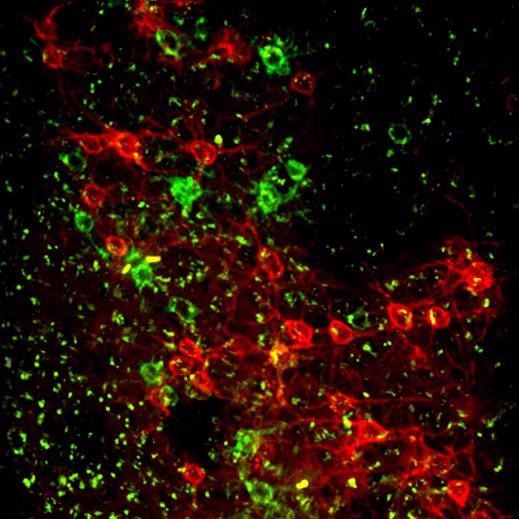
Figure 1. Coronal slice, mouse cortex. Histology of a mouse used for in vivo voltage imaging using Ace-mNeonGreen and Varnam (red) labeled neurons. Both fluorophores express in the cell membrane. Laser source: Fluence Halite (rep. rate 20 MHz, wavelength 1030 nm). Image Credit: Fluence sp. z o.o.
Overcoming Challenges in TPEM
The main challenge in two-photon excitation microscopy is that the likelihood of achieving simultaneous two-photon absorption is extremely low. This means that exceedingly high incident photon densities are necessary in order to produce fluorescence with greater reliability.
As a result, femtosecond lasers are vital tools in TPEM. These devices emit a quick-fire series of extremely short and high-intensity laser pulses to generate the required photon densities without the constant high power output of a continuous laser.
With a heavy dependence on laser performance, the image quality of the two-photon techniques relies primarily on laser parameters. Exact control of pulse duration and minimization of GDD (caused by optical elements in the laser path) are crucial to achieving high peak-power pulses that are as short as possible.
The Halite series of lasers from Fluence Technology can deliver a novel solution to two-photon imaging.4
Low Repetition Rate
Amazingly, a decrease in the rate at which a laser generates pulses (repetition rate) can boost performance. Assuming two lasers have an equivocal average power but different repetition rates, the laser with the lower repetition rate (i.e., fewer pulses per second) must discharge more energy per pulse.
This means that, as a result of lower repetition rates, higher photon densities are produced, thus facilitating higher quality imaging. While competing systems generally offer pulse repetition rates in the region of 80 MHz, the repetition rate of Halite lasers are significantly lower at 20 MHz.
This means that Halite lasers offer a 4x increase in pulse peak power in contrast to an 80 MHz laser with the same nominal power. Figure 2 exhibits a comparison between TPEF conducted using a 20 MHz laser and an 80 MHz laser with an equivalent average power level.
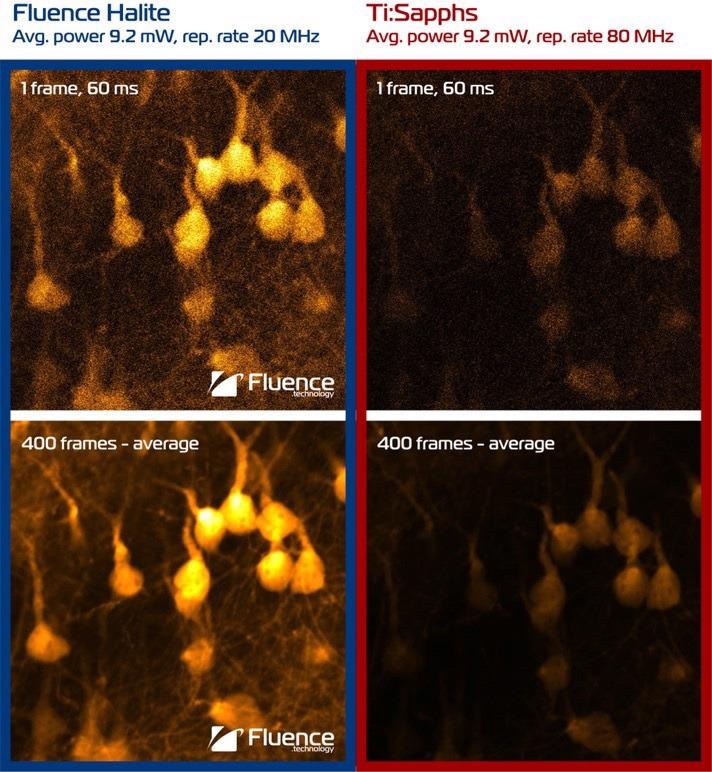
Figure 2. Laser source comparison. Fluence Halite (20 MHz) vs Ti:Sapphire laser (80 MHz). Structural 2-photon imaging at 1030 nm of pyramidal neurons and their dendrites. Fixed tissue, coronal slice, YFP-H mouse line. Image Credit: Fluence sp. z o.o.
It is clear to see that 20 MHz offers a much-improved yield than an 80 MHz laser, thanks to the higher pulse energy.
In the 2 W power class, Halite femtosecond lasers deliver the greatest pulse energy available on the market at up to 100 nJ per pulse. A lower repetition rate also indicates a lower accumulation of heat in the sample, which is vitally important for biological imaging.
All-Fiber Optics Laser systems predicated on crystal technologies (such as Ti:Sapphire lasers) not only come with extremely high capital costs (usually in excess of $150,000) but demand additional maintenance and repair that can have a significant impact on lifetime costs.
The durable and sturdy all-fiber design of the Halite range does away with bulk optics (including saturable absorbers like SESAM) from the optical train, avoiding deterioration of components and eliminating any requirement for future maintenance.
No bulk optics prevent misalignment, limiting the need for time-consuming alignment and optimization. The extraordinary oscillator design of the Halite range guarantees performance in a wide range of conditions that users can depend on: Fluence’s femtosecond oscillator that Halite bases upon demonstrates stable operation even in 40 g shock tests and remains stable across an extensive temperature range.
Halite lasers deliver improved power stability and clean pulses with a standard pulse duration of 200 fs. Meanwhile, built-in GDD precompensation corrects for the group delay dispersion that comes from the microscope’s optics in a range from 10,000 fs2 down to -100,000 fs2.
Each optical component between the laser source and the sample will typically stretch the pulse duration as a result of dispersion, causing an efficiency drop in the process. Automated GDD precompensation tuning may be advantageous for groups assembling microscope prototypes, where an optical train is constantly changing and often unpredictable.
This facilitates the simple addition or subtraction of group delay dispersion to the pulse from the software level so that the optical train of the microscope compresses the pulse duration back to minimal sample value. Thus, automated GDD tuning also enables the user to openly tune with the pulse duration.
Compact Single-Box Design
With an all-in-one compact design, Halite lasers do not necessitate the inclusion of an external cooling unit or controllers. In its class, it is the only laser that is powered from a 24 V power adapter, with all electronics housed in a single laser head.
Halite is not just an elegant solution, but it is also the most compact femtosecond laser for TPEF. Halite lasers are super-reliable light sources that can be smoothly integrated into any two-photon imaging system.
As a result of their compact footprint and stability, they can be easily incorporated into designs of the imaging systems outside the laboratory settings, including hospitals, R&D offices, etc.
Recently, a new generation of Halite has been launched with even more power in the Halite 2, exhibited in figure 3. The new Halite 2 delivers over 2 W of average power and pulse energy as high as >100 nJ, made possible by the reduced base repetition rate down to the level of 20 MHz.
The basic wavelength of 1030 nm in Halite 2 is ideal for TPEF and is also useful for multiphoton excitation fluorescence microscopy MPEF, specifically three-photon microscopy (3PEF).
As well as from the basic wavelength, Halite 2 can also come equipped with the second harmonic option, producing 515 nm laser light at its output, which is optimal for a range of applications such as 2-Photon Polymerization (TPP, 2PP).
To discover more about the Halite range and other high-performance laser products, get in touch with a member of the Fluence Technology team today.
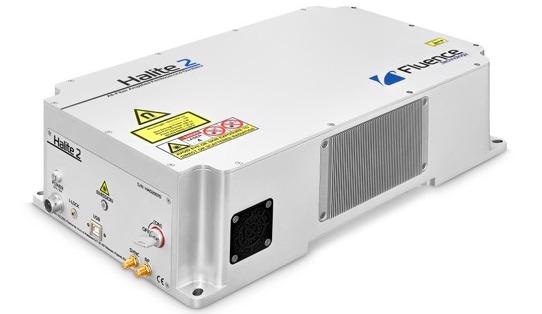
Figure 3. Thanks to its unique technology, Halite 2 is the smallest femtosecond laser with energy level >100 nJ at 20 MHz, and with no external controllers. Image Credit: Fluence sp. z o.o.
References
- Denk, W., Strickler, J. & Webb, W. Two-photon laser scanning fluorescence microscopy. Science 248, 73–76 (1990).
- Larson, A. M. Multiphoton microscopy. Nature Photon 5, 1–1 (2011).
- Helmchen, F., Denk, W. Deep tissue two-photon microscopy. Nat Methods 2, 932–940 (2005). https://doi.org/10.1038/nmeth818.
- Halite: Single-box compact femtosecond laser - Fluence.technology. Fluence.technology. https://fluence.technology/news/introducing-fluence-halite/

This information has been sourced, reviewed and adapted from materials provided by Fluence sp. z o.o.
For more information on this source, please visit Fluence sp. z o.o.