Crystallography provides insight into the structure and properties of materials at the atomic level. While X-ray diffraction has traditionally dominated crystallography, Raman spectroscopy is now emerging as a powerful complementary tool for studying crystalline systems and advancing our understanding of materials.
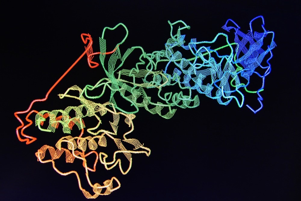
Image Credit: Sergei Drozd/Shutterstock.com
What is Crystallography?
Crystallography holds immense significance due to its profound historical and contemporary contributions to science and technology. It is the science of determining the organization and bonding of atoms within crystalline solids and understanding the structural patterns of crystal lattices.
The discovery of X-rays in 1895 laid the foundation for crystallography. In 1912, German scientists Max Laue, Walter Friedrich, and Paul Knipping's demonstration of X-ray diffraction on crystals provided direct evidence of atomic order, earning Max Laue a Nobel Prize in 1914.
However, the field truly revolutionized in the same year when 22-year-old William Lawrence Bragg revealed how diffraction patterns could determine atomic positions. Working with his father, William Henry Bragg, they rapidly unraveled crystal structures, starting with common salt and diamond, earning them a joint Nobel Prize in 1915.
The Importance of Crystallography
The significance of crystallography lies in its capacity to unveil the secrets of the microscopic world. It allows scientists to visualize and comprehend atomic structures, ranging from natural crystals like diamonds to intricate biological molecules.
Health and medicine have seen the most profound benefits from crystallography, with Dorothy Hodgkin being a standout contributor. She used these methods to decipher the structures of penicillin and insulin, leading to breakthroughs in antibiotics and diabetes treatments. Additionally, the discovery of DNA's double helix structure by James Watson and Francis Crick, aided by Rosalind Franklin's crystallography data, revolutionized genetics.
However, crystallography's impact isn't confined to biology; it is instrumental in understanding material properties and structure, with applications in fields like aerospace engineering, archaeology (e.g., preserving King Henry VIII's ship, the Mary Rose), and the discovery of novel materials such as quasi-crystals and graphene.
Considering that addressing many of the world's challenges hinges on developing new materials and gaining a deeper atomic-level understanding of processes, crystallography will remain a central tool for scientists worldwide in the foreseeable future.
What Technologies Are Used in Crystallography?
X-ray Diffraction
X-ray diffraction was the pioneering technique in crystallography, establishing the foundation for the field. It remains a fundamental and widely employed method for understanding protein structures and functions at the atomic level.
It involves shining X-rays onto a crystal to analyze its diffraction patterns, which provide electron density information for constructing a detailed structural model. Despite its effectiveness, X-ray crystallography has limitations, such as the invisibility of hydrogen atoms and potential damage to biological molecules by X-rays.
Neutron Diffraction
Locating hydrogen atoms is vital for studying enzyme mechanisms and hydrogen bonds in molecules like proteins, DNA, and RNA. Neutron crystallography is a powerful technique for this purpose, revealing protonation states, solvent identities, and hydrogen-related bonds without the risk of damaging the sample.
However, it faces a drawback of low neutron beam flux, requiring large crystals or extended exposure times for smaller crystals to produce measurable diffraction signals.
Nuclear Magnetic Resonance (NMR) Spectroscopy
NMR spectroscopy provides a nuclear site-specific probe of electronic, molecular, and crystal structures, offering more localized information than diffraction methods that depend on long-range molecular ordering in solids.
It complements X-ray diffraction, focusing on nuclei and providing insights into various materials, including non-periodic crystal regions. Recently, researchers have employed two-dimensional NMR spectra to uncover proton-proton contacts and carbon-proton proximities, particularly in cocrystals.
Electron Microscopy
Electron microscopy is well-suited for studying small membrane proteins in their native lipid bilayer environment. Unlike X-ray and NMR techniques, it retains the protein's native state, allowing for time-resolved studies and direct phase information from images.
Where Does Raman Come In?
Raman spectroscopy, a technique based on light scattering, is employed in crystallography to analyze crystalline materials' molecular and atomic structures. It achieves this by measuring the wavelengths of light scattered from a crystal under laser illumination. While most scattered light remains unchanged, a small portion undergoes Raman scattering, producing light at different wavelengths depending on the crystal's chemical structure.
Crystallographers can analyze the resulting Raman spectrum to identify specific molecular bond vibrations, including single bonds (e.g., C-C, N-O, C=C, C-H) and groups of bonds (e.g., in polymer chains, benzene rings, and lattice modes), and gain insights into the crystal's composition and structure.
Raman spectroscopy is a versatile tool used in crystallography to characterize atomic structures, identify polymorphs, investigate crystal phase transformations and extract thermodynamic data, even under extreme conditions such as high pressure, high temperature, or electric/magnetic fields.
Depending on incident light polarization, crystal symmetry, and orientation, Raman scattering is employed to analyze molecular vibrations, offering a "fingerprint" for molecular and crystal structure determination.
Advantages and Limitations of Raman Spectroscopy in Crystallography
Advantages of Raman spectroscopy in crystallography include its non-destructive nature, compatibility with microscopy, and ability to analyze aqueous samples with minimal preparation. It provides valuable chemical information and is suitable for characterizing heterogeneous materials.
However, the limitations of Raman spectroscopy in crystallography stem from its weak signal, susceptibility to fluorescence interference, and surface sensitivity with limited information depth. The use of high-energy lasers can induce sample heating and potential phase changes. While it provides valuable insights, Raman spectroscopy may not offer atomic-scale structural detail, and quantitative analysis can be challenging compared to other techniques.
Recent Research in Raman Crystallography
Understanding Liquid Crystal Phase Transitions through Raman Spectroscopy
In a recent study published in Spectrochimica Acta Part A, scientists used Raman spectroscopy to analyze the structural properties of two high-clearing temperature nematic liquid crystals (LCs). The clearing temperature, pivotal for LCD technology, shifts from ordered liquid crystalline to disordered liquid phase, altering optical properties.
The researchers employed temperature-dependent Raman spectroscopy to investigate these compounds across different temperatures. They found that the Raman spectra's peak positions remained consistent with known bond and functional group assignments, indicating the absence of phase changes or loss of crystallinity in the LC phase.
The deformations observed in the Raman spectra near the clearing temperatures highlighted changes in the LC compounds before reaching the isotropic liquid phase, confirming the presence of a single LC phase.
Studying Catalytic Behavior and Potential Enhancement of Bismuth Molybdate Crystals Using Raman
Bismuth molybdate crystals (Bi2(MoO4)3) are significant due to their versatile properties, making them valuable in catalysis, photocatalysis, and pollutant degradation applications.
A recent study published in Spectrochimica Acta Part A investigated Bi2(MoO4)3 crystals using lattice dynamic calculations and pressure-dependent Raman spectroscopy to reveal structural and vibrational changes under varying pressure conditions, providing insights into their catalytic behavior and potential enhancements for various applications.
The researchers performed lattice dynamics calculations using a rigid ion model, aiding in interpreting the experimental Raman modes observed under normal conditions. Then, they used pressure-dependent Raman spectroscopy to examine Bi2(MoO4)3 crystals in the 20 to 1000 cm−1 spectral range under pressures from 0.1 to 14.7 GPa. The Raman spectra showed significant changes at 9.2, 4.9, and 2.6 GPa, indicating structural phase shifts.
This research helps expand our knowledge of how Bismuth molybdate behaves under extreme conditions, which can have implications for its practical applications.
Concluding Remarks
Raman spectroscopy is a non-destructive and versatile technique in crystallography, offering valuable insights into crystal phases, molecular vibrations, and chemical composition. Its strengths and limitations make it a valuable tool for characterizing crystalline materials, especially when combined with other analytical methods for a comprehensive analysis.
More from AZoOptics: Raman Spectroscopy Market Trends: A Worldwide Overview
References and Further Reading
Basumatary, J., Gangopadhyay, D., Nath, A., & Thingujam, K. D. (2023). Studies of temperature dependent Raman spectroscopy of two nematic liquid crystalline compounds of homologous series. Spectrochimica Acta Part A: Molecular and Biomolecular Spectroscopy, 300, 122898. https://doi.org/10.1016/j.saa.2023.122898
Blakeley, M. P., Langan, P., Niimura, N., & Podjarny, A. (2008). Neutron crystallography: opportunities, challenges, and limitations. Current opinion in structural biology, 18(5), 593-600. https://doi.org/10.1016/j.sbi.2008.06.009
Bryce, D. L. (2017). NMR crystallography: structure and properties of materials from solid-state nuclear magnetic resonance observables. IUCrJ, 4(4), 350-359. https://doi.org/10.1107/S2052252517006042
Hite, R. K., Schenk, A. D., Li, Z., Cheng, Y., & Walz, T. (2010). Collecting electron crystallographic data of two-dimensional protein crystals. In Methods in enzymology (Vol. 481, pp. 251-282). Academic Press. https://doi.org/10.1016/S0076-6879(10)81011-0
Horiba. (2023). What is Raman Spectroscopy? [Online]. Available at: https://www.horiba.com/int/scientific/technologies/raman-imaging-and-spectroscopy/raman-spectroscopy/
Institute of Physics. (2023). Crystallography. [Online]. Available at: https://www.iop.org/explore-physics/big-ideas-physics/crystallography
Krylov, A. (2020). Raman Spectroscopy of Crystals. Crystals, 10(11), 981. https://doi.org/10.3390/cryst10110981
Martineau, C., Senker, J., & Taulelle, F. (2014). NMR crystallography. Annual Reports on NMR Spectroscopy, 82, 1-57. https://doi.org/10.1016/B978-0-12-800184-4.00001-1
Saraiva, G. D., de Castro, A. R., Teixeira, A. M. R., Neto, V. S., Lima Jr, J. A., Juca, R. F., ... & Paraguassu, W. (2023). Pressure-dependence Raman spectroscopy and the lattice dynamic calculations of Bi2 (MoO4) 3 crystal. Spectrochimica Acta Part A: Molecular and Biomolecular Spectroscopy, 297, 122711. https://doi.org/10.1016/j.saa.2023.122711
Singh, D. (2020). Introductory Chapter: Electron Crystallography. IntechOpen. https://doi.org/10.5772/intechopen.93063
Tuschel, D. (2012). Raman Crystallography. [Online]. Available at: https://www.spectroscopyonline.com/view/raman-crystallography
Uddin, Y. M., & Schmidt-Krey, I. (2015). Inducing two-dimensional crystallization of membrane proteins by dialysis for electron crystallography. In Methods in Enzymology (Vol. 557, pp. 351-362). Academic Press. https://doi.org/10.1016/bs.mie.2014.12.022
Disclaimer: The views expressed here are those of the author expressed in their private capacity and do not necessarily represent the views of AZoM.com Limited T/A AZoNetwork the owner and operator of this website. This disclaimer forms part of the Terms and conditions of use of this website.