The compromise between signal strength and fluorescence background is often the driving factor when considering excitation wavelength for Raman spectroscopy. But what if fluorescence background could be completely eradicated?
UV Raman facilitates just that. The article examines its advantages, the technical difficulties it has overcome, and some fascinating applications. Throughout, UV resonance Raman (UVRR) is also detailed, a methodology that can produce considerable signal enhancement and improved analytical specificity under the right circumstances.
Avoiding the Fluorescence Window
The effective efficiency of Raman scattering scales contrary to excitation wavelength to the fourth power (1/λex4), which as such presents a compelling case for UV excitation. Theoretically, this means that excitation with UV light at 248 nm can yield signals >20x stronger than at 532 nm, or 100x greater than at 785 nm. However, in practice, there is a more persuasive reason for UV Raman excitation: eradicating background fluorescence.
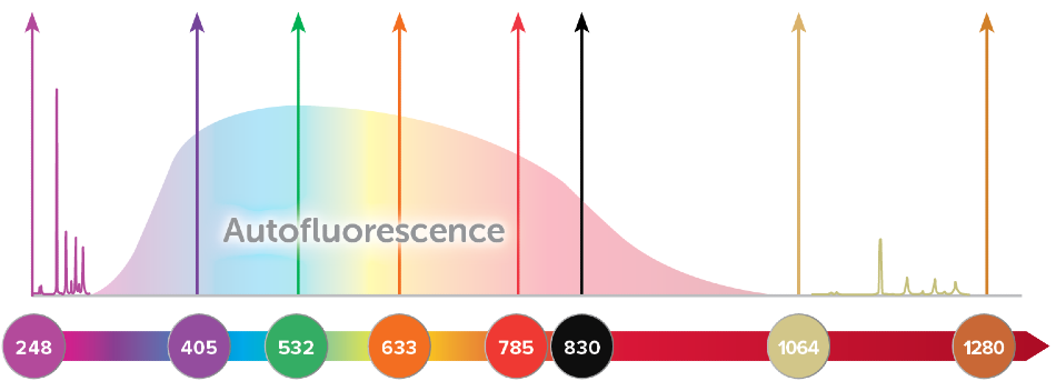
Numerous Raman active molecules also demonstrate fluorescence when excited with a laser, leading to a broad background that is typically intensified several orders of magnitude more than the Raman signal. This broad emission may converge with the complete Raman spectrum when visible light is used. This may reduce the signal-to-noise ratio of the Raman spectrum or completely obscure it.
When the molecular structure is complex, the fluorescence background is greatest. This is observed in organic compounds and biological samples but can also take place if fluorescent impurities are present in the sample.
The reduction of fluorescence can be achieved by using a longer wavelength like 785 or 830 nm. Furthermore, elimination of fluorescence is almost completely possible using 1064 nm, though some biological samples still undergo undesirable heating when excited with 1064 nm Raman.
Alternatively, exciting and capturing Raman spectra at wavelengths below the fluorescence window, using UV Raman, is another option to help mitigate fluorescence. Generally, fluorescence occurs at wavelengths longer than 300 nm, therefore, by utilizing an excitation laser at or below 266 nm, a complete 4000 cm1 spectral range covering the fingerprint and functional bands can be collected rather easily with little to no interfering background. Furthermore, the use of an excitation source in the UV range may lead to a large resonant improvement of the Raman signal for some samples: this method is known as resonance Raman spectroscopy.
UV Resonance Raman (UVRR)
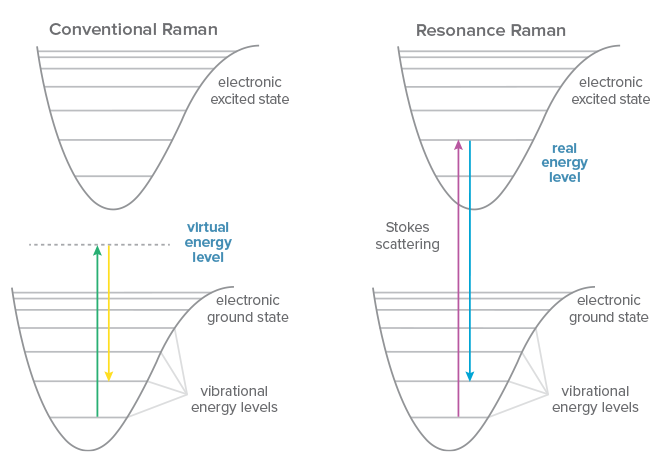
Traditional Raman scattering is an extremely weak phenomenon and one that takes place to some degree irrespective of the excitation wavelength used. However, if the energy of the excitation laser happens to correspond with an electronic transition within the molecule under scrutiny, the signal could be improved by a factor of 102-106: a method termed resonance Raman spectroscopy.
Even excitation near the electronic transition of a molecule can generate ‘pre-resonance’, yielding 5-10x more signal. Resonance Raman enables rapid measurements with a greater signal to noise ratio and allows measurement of reduced concentrations or even trace detection.
Another advantage of resonance Raman is greater selectivity because the signal enhancement only occurs for electronic transitions that correspond with the laser wavelength. This can enable favored excitation of a molecule within an intricate sample matrix, or selective enhancement of the signal from a particular subgroup within a macromolecule.
UV resonance Raman (UVRR) utilizes the resonance of UV light with chromophores and aromatics in intricate biomolecules to examine the structure and dynamics of proteins and nucleic acids, from folding to interactions and alterations in the environment. The selection of wavelength resolves the specific structure for which resonance occurs, amplifying its signal selectively to generate a comparatively simple Raman spectrum despite the complex sample.
Advances in UV Raman Instrumentation
The short wavelength of UV light demands more from the instrumentation used for Raman scattering. While the selection may be restricted, commercially available components now make UV Raman and UVRR accessible for a much wider range of applications than ever before. Affordable, compact UV lasers with greater lifespan and power output have led to a decrease in the size of UV Raman systems. They include the two most desirable options, NeCu hollow-cathode metal-ion lasers at 248.6 nm and quadrupled, diode-pumped Nd:YAG lasers at 266 nm.
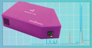
Introducing the first compact, modular UV Raman spectrometer, the WP 248 from Wasatch Photonics. It covers the range 400-3200 cm-1 with 14 cm-1 resolution and is designed for use with a 248.6 nm NeCu laser. Image Credit: Wasatch Photonics, Inc.
On the detection side, the spectrometer being utilized must be both high-resolution and high-throughput. Only ~27 nm of the spectral range is needed to generate a 4000 cm-1 Raman spectrum if the excitation laser is 248 nm; 0.1 nm resolution is equal to 16 cm-1. This calls for the long-pass filter used for rejection of Rayleigh scatter to be much steeper than its visible counterparts.
Thus, to acquire enough signal in reasonable measurement time, considering the narrow spectral range in the UV, the spectrometer demands extremely high throughput. This can be accomplished using efficient optics, a high numerical aperture input, and a sensitive detector. Sensitive detection of UV Raman is crucial to maintain short exposure times and significantly reduces potential damage to delicate samples, such as biological materials.
Applications of UV Raman
Exploration of UV Raman has been performed for many applications, only some of which harness the additional benefits of UV resonance Raman. In the field of gas measurement, testing of UV Raman has shown it to be a viable technique for pressure measurement1 and for the trace detection of nitrogen2. It has also been put to use in combustion studies3 and for analysis of fuel/air mixing4.
As a tool for analyzing solid-state materials, it can provide greater insight into structural, optical, and electronic properties of materials manufactured for electronics5. In the field of diamond growth, UV Raman can help identify and evaluate impurities that would strongly exhibit fluorescence with visible Raman6.
The restraining of fluorescence background in combination with a resonant enhancement of signal provides UV Raman with an advantage for trace detection of analytes within intricate samples. These range from detecting cocaine in saliva7 to the presence of environmental pollutants such as carcinogenic PAHs.6 Even the sheer reduction in the background has proven to aid the characterization of strongly fluorescing, colored food samples including dark beverages and edible oils8.
UV Raman is also the favored method for the standoff detection of explosive threats and chemical warfare agents by security forces. It should be noted that this is not an application for a research-grade spectrometer, as these systems demand high-power lasers and very specific optics to accomplish the long-working distance required.9,10
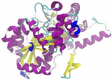
However, UV resonance Raman has made its greatest impact as a viable method for probing molecular structure and dynamics in biological systems. It permits the particular excitation of a specific, localized segment of interest within a macromolecule, such as an amino acid or nucleic acid. In doing so, it improves the signal from only that substructure to supply comparatively clean Raman spectra from within highly complex samples. This makes it priceless concerning the studies of protein structure and folding6, as well as the influence of environment or interactions with other molecules.11,12
Therefore, UVRR could also become an extremely useful monitoring tool in bioprocessing.13 It has even been investigated for use in monitoring the mode of action of antibiotics for specific antibiotic/bacterium pairs14.
(For additional details on these applications, explore the UV Raman reference list below.)
Conclusion
Progress in the technologies necessary to utilize UV Raman, such as compact lasers and the new WP 248 Raman spectrometer has contributed to this powerful technique becoming increasingly available. Furthermore, now more than ever UVRR is appropriate for a broad range of applications from materials analysis and trace detection to studies of biological structure and dynamics.
Including UV in your suite of Raman instrumentation presents the opportunity to acquire fluorescence-free spectra and the potential for improving signal through UVRR for some samples, thus complementing the broad spectrum of visible and NIR wavelengths now available for Raman spectroscopy.
References and Further Reading
- Gu, Y., et al. “Pressure dependence of vibrational Raman scattering of narrow-band, 248-nm, laser light by H2, N2, O2, CO2, CH4, C2H6, and C3H8 as high as 97 bar.” Applied Physics B 71.6 (2000): 865-871.
- Hargis, P. J. “Trace detection of N 2 by KrF-laser-excited spontaneous Raman spectroscopy.” Applied optics 20.1 (1981): 149-152.
- Raffius, Thomas, et al. “Flame-temperature, light-attenuation, and CO measurements by spontaneous Raman scattering in non-sooting diesel-like jets.” Combustion and Flame 176 (2017): 104-116.
- Grady, Nathan R., et al. “Raman scattering measurements of mixing and finite-rate chemistry in a supersonic reacting flow over a piloted, ramped cavity.” Combustion and Flame 165 (2016): 310-320.
- Liu, Hsiang-Lin, et al. “Deep-ultraviolet Raman scattering spectroscopy of monolayer WS 2.” Scientific reports 8.1 (2018): 1-10.
- Asher, Sanford A., Calum H. Munro, and Zhenhuan Chi. “UV lasers revolutionize Raman spectroscopy.” Laser Focus World 33.7 (1997): 99-109.
- D’Elia, Valentina, et al. “Ultraviolet resonance Raman spectroscopy for the detection of cocaine in oral fluid.” Spectrochimica Acta Part A: Molecular and Biomolecular Spectroscopy 188 (2018): 338-340.
- El-Abassy, Rasha M., Bernd von der Kammer, and Arnulf Materny. “UV Raman spectroscopy for the characterization of strongly fluorescing beverages.” LWT-Food Science and Technology 64.1 (2015): 56-60.
- Nagli, L., et al. “Absolute Raman cross-sections of some explosives: Trend to UV.” Optical Materials 30.11 (2008): 1747-1754.
- Choi, Sun‐Kyung, et al. “Analysis of Raman Spectral Characteristics of Chemical Warfare Agents by Using 248‐nm UV Raman Spectroscopy.” Bulletin of the Korean Chemical Society 40.3 (2019): 279-284.
- Chinsky, L., et al. “Resonance Raman spectra of poly (l‐lysine), aromatic amino acids, l‐histidine and native and thermally unfolded ribonuclease A.” Journal of Raman spectroscopy 16.4 (1985): 235-241.
- Jakubek, Ryan S., et al. “Ultraviolet resonance Raman spectroscopic markers for protein structure and dynamics.” TrAC Trends in Analytical Chemistry 103 (2018): 223-229.
- Goodacre, Royston, and Lorna Ashton. “Application of deep UV resonance Raman spectroscopy to bioprocessing.” European Pharmaceutical Review 16.3 (2011): 46-49.
- López-Díez, E. Consuelo, et al. “Monitoring the mode of action of antibiotics using Raman spectroscopy: investigating subinhibitory effects of amikacin on Pseudomonas a eruginosa.” Analytical chemistry 77.9 (2005): 2901-2906.
- Sapers, Haley M., et al. “Using Deep UV Raman Spectroscopy to Identify In Situ Microbial Activity.” AGUFM 2017 (2017): B11G-1744.

This information has been sourced, reviewed and adapted from materials provided by Wasatch Photonics, Inc.
For more information on this source, please visit Wasatch Photonics, Inc.