Heme, which is the precursor to hemoglobin, consists of an organic molecule known as porphyrin that is attached to an iron atom. The iron atom of heme reversibly binds to an oxygen atom during the oxygenation of blood in the lungs. In most vertebral organisms, the amount of oxygen available to the tissues directly correlates with the color of blood as it circulates throughout the vascular system. As a result, many medical professionals throughout history have relied upon the color of blood to determine certain health characteristics of the patient.
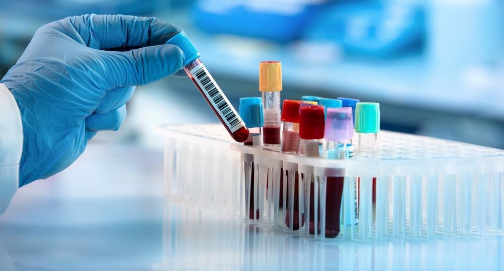
Image Credit: angellodeco/Shutterstock.com
The Optical Properties of Blood
Aside from subjectively monitoring the optical properties of blood, several technologies have been developed to gain insight into the optical absorption behavior of blood and assess the oxidation state of iron within heme molecules. To this end, experimental X-ray diffraction, optical spectroscopy, and several quantum chemistry approaches have been applied.
With specific regards to hemoglobin, experimental optical spectroscopy studies have described three forms, including oxy-Hb, deoxy-Hb, and met-Hb. However, there remains a lack of information on the optical-electronic transition properties of the main hemoglobins that can be used to determine the color of blood.
Theoretical Predictions of Hemoglobin Structures
In a recent Royal Society of Chemistry study, researchers sought to utilize Raman spectroscopy to gain insight into the optical properties of blood samples at the cellular level.
Initially, the researchers utilized density functional theory (DT) to reproduce the optical absorption and circular dichroism behavior of met-Hb, deoxy-Hb, and oxy-Hb. This was followed by calculations of the optical absorption and natural transition orbitals (NTO) of these three forms of hemoglobin.
Notably, the spin densities and atomic charges calculated for met-Hb, deoxy-Hb, and oxy-Hb were all similar to their previous assignments; however, the atomic charge of iron in met-Hb was found to be slightly less than anticipated. Upon comparison of the orbital components of heme, the researchers observed considerable differences in the electronic elements specific to met-Hb, deoxy-Hb, and oxy-Hb as compared to previous reports.
These theoretical predictions were then compared to the experimental visible (VIS) absorption and circular dichroism (CD) spectra of three types of hemoglobin.
Several ligand to metal charge transitions reflected the scarlet red color often associated with oxygenated blood in vertebrates, whereas the charge-transfer transition in deoxy-Hb from the binding orbital of porphyrin to iron appears to be responsible for the dark purple color of blood in veins.
The computational studies described here provided an accurate representation of the electronic transitions that occur within blood. The researchers successfully correlated these reactions to the different appearances that blood can have, depending upon the type of hemoglobin present in a given sample.
A New Approach to Optical Blood Analysis
Although frequency-resolve Raman spectroscopy has been successfully applied to the biochemical analysis of blood cells at the sub-cellular level, the utilization of both resonant and non-resonant Raman spectroscopy approaches has not been applied to the analysis of blood samples.
To this end, the researchers acquired pre-resonant experimental Raman spectra for oxy-Hb, deoxy-Hb, and met-Hb. These results were then compared with theoretical Raman resonances, by subtracting the spectrum of oxy-Hb by deoxy-Hb alone and with contributions of met-Hb.
The researchers found that the pre-resonant Raman spectra results were consistent with the calculated dispersions for all three forms of hemoglobin.
Raman microscopy was then used to analyze and compare the properties of both discocyte (healthy) and echinocyte (abnormal) blood cells. Within the healthy discocyte sample, the researchers observed that deoxy-Hb has a tendency to be displaced from its central regions and the side of the cell. At these sample locations, oxy-Hb is more likely to be abundant.
Comparatively, within the echinocyte blood sample, deoxy-Hb was more likely to occupy the central region of the cells, thereby demonstrating a disruption in the distribution of both oxy-Hb and deoxy-Hb in abnormal blood cells.
These observations correlate with previous reports on the increased height of the center of echinocytes, as well as the formation of spicules, which are cytoskeletal rearrangements that are characteristic of echinocytes. Thus, the researchers hypothesize that spicules may actually release hemoglobins from outside the cell, which is a phenomenon that has been reported in hemolytic anemias with echinocytosis.
The researchers also utilized Raman spectroscopy to assess the function of met-Hb in the shape of both normal and abnormal blood cells. The researchers found that the enzyme cytochrome-b5-MHb reductase plays an important role in maintaining the center of the toroidal channel within the discocyte.
Although this enzyme appears to be active in the center of the echinocyte as well, it is unable to prevent the accumulation of met-Hb within the spicules present in these abnormal cells, thus impacting their shape.
Study Takeaways
The researchers found that Raman microscopy can be used to acquire information on blood biochemical processes at the cellular level by specifically monitoring the redistributions of three different forms of hemoglobin.
Since this type of analysis would not be easy to achieve with other microscopic techniques, the Raman approach described here has the potential to assist in the diagnosis of various hematological conditions such as blood oxidative stress, methemoglobinemia, and other anemic tendencies.
References and Further Reading
Volkov, V. V., McMaster, J., Aizenberg, J., & Perry, C. C> (2022). Mapping blood biochemistry by Raman spectroscopy at the cellular level. Royal Society of Chemistry 13;133-140. doi:10.1039/D1SC05764B.
Wood, B. R., Kochan, K., & Marzec, K. M. (2020). Chapter 13 – Resonance Raman spectroscopy of hemoglobin in red blood cells. Vibrational Spectroscopy in Protein Research; 375-414. doi:10.1016/B978-0-12-818610-7.00013-X.
Disclaimer: The views expressed here are those of the author expressed in their private capacity and do not necessarily represent the views of AZoM.com Limited T/A AZoNetwork the owner and operator of this website. This disclaimer forms part of the Terms and conditions of use of this website.