Reviewed by Frances BriggsJul 31 2025Reviewed
Researchers unveil a pocket-sized scanner that uses AI and dual imaging to detect skin cancer noninvasively, offering faster, more accurate diagnoses.
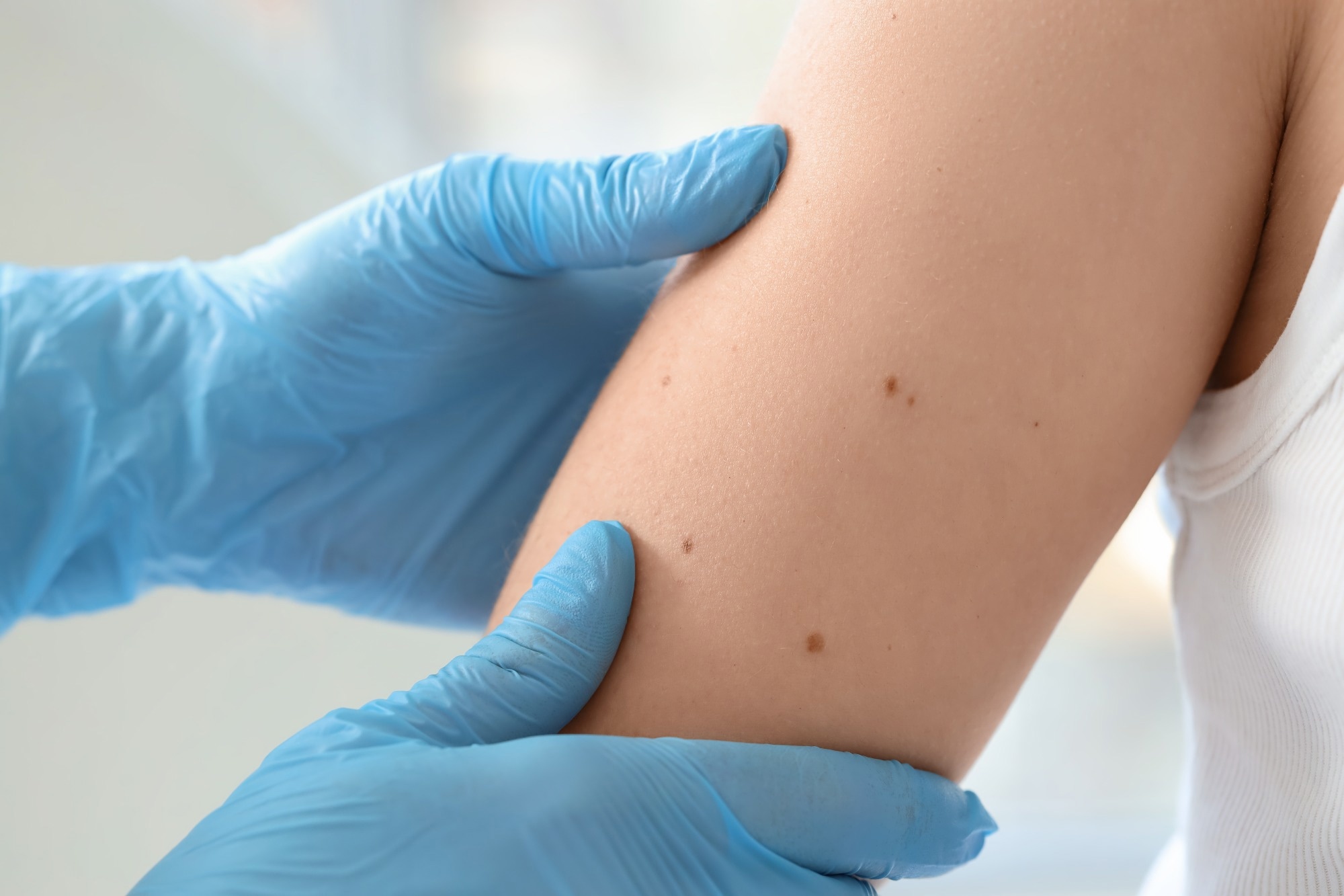
Image Credit: /Shutterstock.com
In a study published in the Journal of Biomedical Optics, a team has introduced a compact imaging device that merges two sophisticated technologies to examine the structure and chemical composition of skin cancers.
Skin cancer is the most prevalent type of cancer in the world, and effective treatment is dependent on early detection. Designed for clinical use, this scanner could streamline diagnosis and treatment decisions, while sparing patients from the scalpel.
The researchers have demonstrated their small, noninvasive imaging device that combines line-field confocal optical coherence tomography (LC-OCT), which captures cellular-level images of tissue, with confocal Raman microspectroscopy, a technique that maps the chemical makeup of targeted areas. By integrating these modalities, not only can clinicians spot abnormal structures, but also analyze their biochemical signatures.
The system analyzed over 330 skin cancer samples in the course of a year. The clinical evaluation focused primarily on nonmelanoma types such as basal cell carcinoma and squamous cell carcinoma.
Researchers used LC-OCT to identify suspicious formations and then employed Raman spectroscopy to gather over 1,300 chemical spectra from those regions. To assess the data, they developed an AI algorithm to spot patterns found in cancerous tissues.
The model proved highly effective, showing a classification accuracy of 95 % for basal cell carcinoma and 92 % when both types of cancer were included. These findings indicate that the technology could accurately identify cancerous structures based on their chemical fingerprints.
The data also revealed clear chemical distinctions between cancer forms, offering new information on their biological differences.
This dual-imaging method could result in more precise, less invasive skin cancer diagnosis in the future. Combining structural and chemical information in this way may allow doctors to make faster and more informed treatment decisions, thereby improving patient outcomes.
Journal Reference:
Ayadh, M., et al. (2025). AI-assisted identification of nonmelanoma skin cancer structures based on combined line-field confocal optical coherence tomography and confocal Raman microspectroscopy. Journal of Biomedical Optics. doi.org/10.1117/1.JBO.30.7.076008.