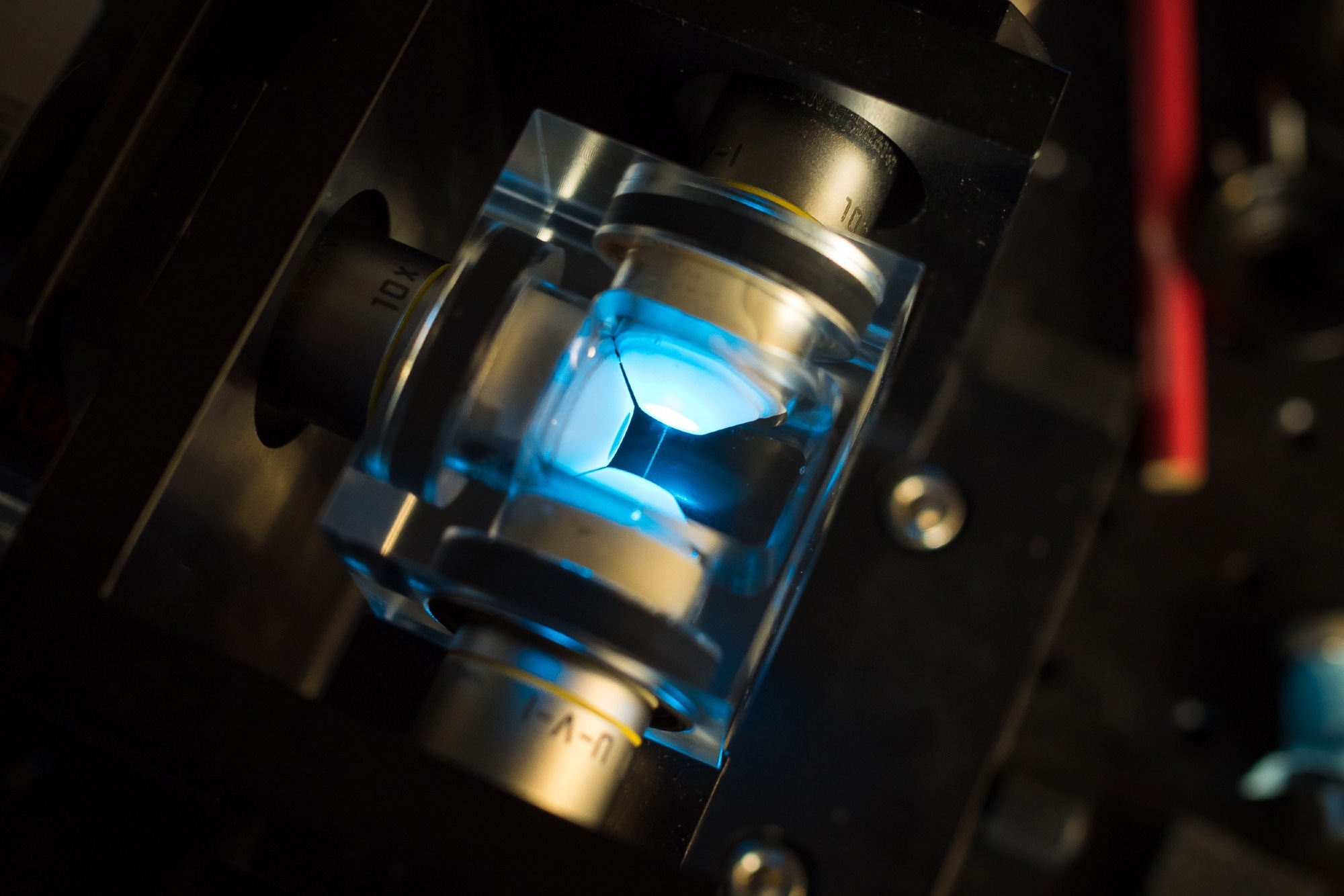
A recent study in Nature Photonics introduced a new computational technique that significantly enhances laser scanning microscopy: super-resolution sectioning image scanning microscopy (s2ISM).
This method aims to enable simultaneous super-resolution and optical sectioning to address the limitations of imaging thick biological samples.

Image Credit: Micha Weber/Shutterstock.com
The Challenge of Optical Sectioning and Resolution
Optical microscopy is an important technique for 3D imaging of biological structures. However, conventional wide-field systems struggle with optical sectioning due to the “missing cone” effect, which causes out-of-focus blur and reduces contrast and resolution.
Techniques like selective plane illumination and multiphoton excitation minimize out-of-focus regions, while confocal laser scanning microscopy (CLSM) improves resolution using a pinhole to block unwanted fluorescence. But, achieving super-resolution often requires a nearly closed pinhole, significantly reducing the signal-to-noise ratio (SNR).
Image scanning microscopy (ISM) uses a detector array to enhance CLSM, such as a single-photon avalanche diode (SPAD) array, where each element acts as a small pinhole, improving light collection and resolution. Despite advancements like these, existing ISM reconstruction techniques struggle to maintain high SNR and effective optical sectioning.
Introducing s2ISM: A Novel Computational Framework
In this study, researchers introduced s2ISM, a computational reconstruction method that uses ISM datasets without requiring hardware modifications. They built a custom ISM microscope with a 5×5 asynchronous-readout SPAD array and software for precise data acquisition.
s2ISM interprets the ISM data as a set of confocal-like images, each blurred by a unique point spread function (PSF) based on the detector element’s position and the emitter’s axial depth. It simplifies computation by modeling image formation with in-focus and out-of-focus planes, effectively separating the background from the signal.
Using a maximum likelihood approach, similar to Richardson-Lucy deconvolution, s²ISM iteratively reconstructs two images from a single-plane ISM dataset: one with enhanced resolution and optical sectioning and another representing background.
This process preserves the focal plane signal and improves SNR without compromising on resolution.
The authors also developed data-driven strategies to estimate essential parameters directly from the raw data, which eliminated the need for further calibration with sub-diffraction sources.
Demonstrated Advantages: Resolution, and Signal Quality
The study validated s2ISM using simulations and experimental imaging of biological samples and resolution targets. In comparison to confocal microscopy and reconstruction methods, such as adaptive pixel reassignment (APR) and multi-image deconvolution, s²ISM consistently provided superior lateral resolution, optical sectioning, and SNR.
Using fluorescent resolution targets with line spacings down to 0 nm, this super-resolution technique resolved details beyond the diffraction limit, outperforming APR and confocal approaches.
Imaging a 3D stair of ring-shaped structures, axially spaced by 250 nm, demonstrated that s2ISM doubled the optical sectioning performance of conventional ISM techniques, effectively rejecting out-of-focus light without relying on assumptions about sample structure.
s2ISM enabled digital super-resolution by leveraging spatial shifts in scanned images, allowing for under-sampled acquisition with pixel sizes up to twice the Nyquist limit. This supports faster imaging, making it ideal for live-cell studies.
Its versatility was demonstrated across multicolor fluorescence imaging, live-cell mitochondrial dynamics, and deep tissue imaging with two-photon excitation, improving resolution and background rejection.
The study also extended the method to fluorescence lifetime imaging microscopy (FLIM) as s2FLISM, incorporating temporal data to distinguish in-focus and out-of-focus lifetimes. This allowed more accurate and robust lifetime estimation, particularly for fluorophores with overlapping emission spectra but distinct lifetimes.
Versatility Across Imaging Modalities and Sample Types
The s2ISM framework is versatile: it is compatible with any laser scanning microscope using a detector array and incoherent light detection, working with fluorescence, Raman, and photothermal imaging. Its ability to enhance resolution and optical sectioning without hardware modifications supports its adoption in biological and biomedical research.
Pairing this method with wavefront sensing and aberration correction could also improve imaging depth in scattering tissues.
The novel technique also demonstrated effectiveness in live-cell time-lapse imaging and deep tissue studies. In HeLa cells, it preserved high resolution and optical sectioning across time-lapse acquisitions, maintaining spectral fidelity while enhancing contrast and spatial detail.
The framework improved deep imaging performance, as shown in ~10 μm-deep Purkinje cell images from mouse cerebellum slices.
Download your PDF copy now!
Advancing Microscopy: Toward Enhancing 3D Imaging
Significant advancements may be possible using s2ISM in laser scanning microscopy. It enables simultaneous super-resolution and optical-sectioning without increasing system complexity or compromising image quality.
As a computational method, it can be integrated into existing LSM systems with detector arrays, enhancing accessibility and adoption.
By fully utilizing structured detection, s2ISM delivered high-resolution, optically sectioned images across a wide range of biological samples. Using open-source implementation and data-driven parameter estimation, it supported reproducibility and broad applicability.
Compatible with live-cell imaging, deep tissue studies, and time-resolved modalities like FLIM, this technique may become a foundational tool in microscopy and in the rapidly evolving field of high-resolution optical imaging.
Journal Reference
Zunino, A., et al. Structured detection for simultaneous super-resolution and optical sectioning in laser scanning microscopy. Nat. Photon. (2025). DOI: 10.1038/s41566-025-01695-0, https://www.nature.com/articles/s41566-025-01695-0
Disclaimer: The views expressed here are those of the author expressed in their private capacity and do not necessarily represent the views of AZoM.com Limited T/A AZoNetwork the owner and operator of this website. This disclaimer forms part of the Terms and conditions of use of this website.