Apr 19 2018
A team of scientists from Ludwig-Maximilians-Universität (LMU), the Max Planck Institute of Quantum Optics (MPQ) and the Technical University of Munich (TUM) have embarked on a mission towards the clinical application of a new laser-based source of X-rays.
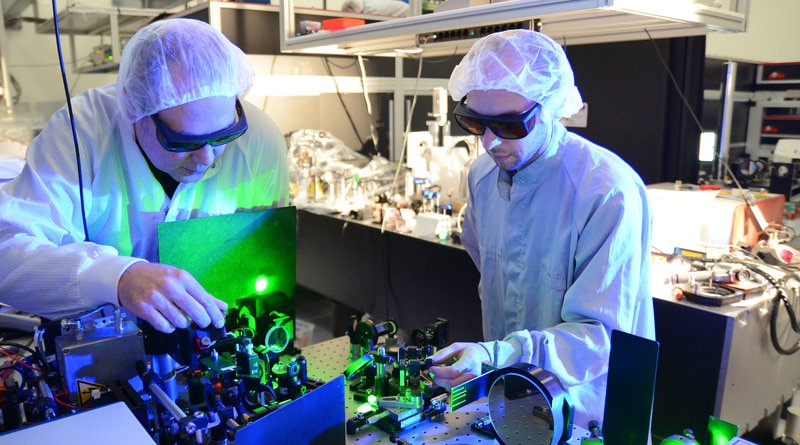 The further development of ATLAS, the high-performance laser in LMU’s Laboratory for Extreme Photonics, paved the way for the tomographic reconstruction of the three-dimensional fine structure of a bone sample within a few minutes. (Image credit: Thorsten Naeser)
In recent times, they proved that the instrument enables the tomographic reconstruction of the 3D fine structure of a bone sample in a matter of a few minutes. Until now, laser-based measurements of this kind took a number of hours. The innovation was made possible by the further progress of ATLAS, the high-performance laser in LMU’s Laboratory for Extreme Photonics (LEX Photonics) der LMU on the Research Campus in Garching. Reconstruction of the sample from the imaging data was also enabled by the use of specifically designed computer programs.
The X-rays used to check the contents of passengers’ baggage at airports or for medical imaging are produced by X-ray tubes, whose design has stayed fundamentally unchanged for more than a century. Research scientists like to use what is known as synchrotron radiation as an X-ray source. Synchrotron radiation is many times brighter and therefore allows one to perform far more thorough structural analyses. However, sources of synchrotron radiation are comparatively thin on the ground, as its generation necessitates the acceleration of electrons to ultrarelativistic velocities (speeds nearer to that of light), and construction of particle accelerators of the required size is massively expensive.
To harness the benefits of synchrotron radiation for basic use in medicine, physicists at LMU, the MPQ, and the TUM have been studying the application of high-performance lasers to the creation of X-rays. In their system, hydrogen atoms are irradiated with very intense pulses of laser light. The extremely energetic optical fields strip the electrons from the atoms and part of the ionized plasma electrons are accelerated. Concurrently, these electrons oscillate in the plasma fields, which cause them to produce the desired synchrotron radiation, i.e. high-intensity X-rays. Furthermore, this entire process occurs over a path-length of less than 15 mm. Thus laser-based X-ray sources have a much smaller footprint, and are a lot less costly to build, than conventional synchrotrons, but create X-radiation of similar quality.
In the initial trials undertaken at the Max Planck Institute in 2015, the research team was able to derive the 3D structure of an insect from 2D projection images captured from various angles. For the latest experiments, done in the Laboratory for Extreme Photonics, Prof. Stefan Karsch and his colleagues have increased the pulse rate, photon yield, and photon energies, and this time they decided to image a sample of human bone. As a result of an enhanced processing algorithm, developed by Prof. Franz Pfeiffer and his team at the TUM, the team needed to gather considerably less data than before. Consequently, the full tomogram could be attained within less than three minutes.
The project was conceived and started in the Munich-Centre for Advanced Photonics (a Cluster of Excellence) and is undergoing additional development at the Center for Advanced Laser Applications (CALA) in Garching. The laser systems available at CALA are projected to greatly improve the efficiency of the source and the quality of the radiation produced, thereby making this new form of tomography accessible for clinical applications for the first time.