Professor Richard Weston, a renowned architect, lecturer and author of numerous books on architecture and mineralogy, has harnessed the latter interest to form a company, Richard Weston Studio, specialising in creating aesthetic designs and artworks from small specimens of minerals, fossils and stones. In 2011 he was described by a UK newspaper, the Independent on Sunday, as "the breakout star of Britain's Next Big Thing", a BBC2 TV series in which the buying teams of three high street giants asked the public to supply them with the next bestselling products.
His success stemmed from a series of images of natural materials he captured using a high resolution document scanner to produce designs for a collection of silk scarves. Introduced in 2010 to the shelves of the Liberty department store in London, the scarves quickly became one of the shop’s best-selling lines. Two years later, four of Prof Weston’s photographs enlarged to five metres by three metres graced the entrance to accommodation in the Olympic Village in Stratford during the London Olympic Games.
Outside the fashion industry, his high resolution digital images and 3D models offer a vast range of possibilities in architecture and the design of urban spaces, gardens and interiors, from printing or etching tiles and digitally weaving rugs to decorating a wall or an entire building. The work has led to increasing international recognition, with recent magazine articles appearing in the USA, The Ukraine and The Netherlands and two books on textile design published in the UK.
Current work at the studio (www.richardwestonstudio.com) is a far cry from early attempts to capture decent pictures of a £143 ammonite specimen Prof Weston bought in 2003 from a local crystal shop in Cardiff, south Wales. He recalled, “My scanner cost less than the ammonite and resulted in a disappointing, muddy brown picture. So I invested in an Epson 3,200 dpi A3 scanner and that produced a beautiful result.
“As the years progressed, I bought many mineral specimens from around the country and online, some of which were very small. Whereas a 20 mm square is good enough for the Epson scanner to register a decent image of high enough resolution to be enlarged, I sometimes wanted to concentrate on details of samples as small as 1 mm square.
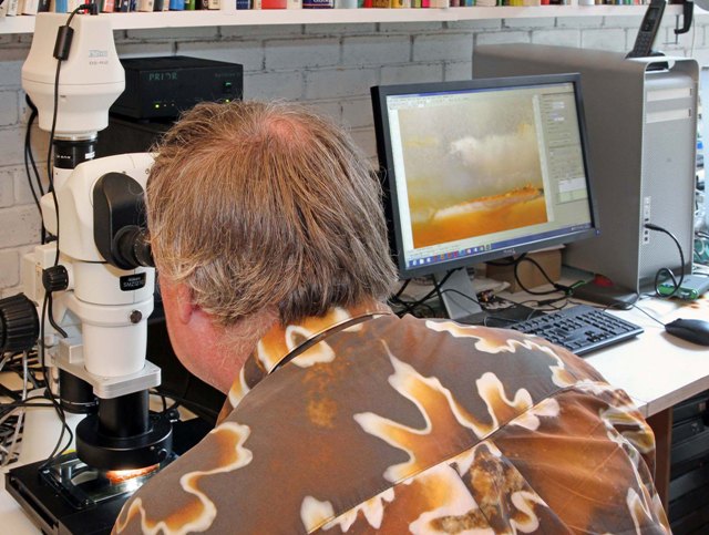
“A conversation with a colleague at Cardiff University working on a medical application convinced me that a photomicrography station was the way to go. As a Nikon Metrology microscope was in use there, I decided to opt for the same supplier.”
In September 2014, he bought an SMZ1270 stereoscopic microscope equipped with a Prior moving stage and a Z-axis drive, both with a positioning accuracy of one micron, a Nikon camera and sophisticated NIS-Elements frame selection and stitching software, all supplied by Nikon Metrology (www.nikonmetrology.com). The scope of projects that could be carried out increased dramatically and so did the quality of the images produced.
The instrument as originally delivered had a x65 zoom lens and a digital camera capable of taking 12 MB TIFF files per frame. It was so successful that it was upgraded within two months to include a x200 zoom lens, enhanced lighting options and a 45 MB per frame Nikon DS-Ri2 camera with full-frame CMOS sensor. The PC-to-camera control unit inside the camera head enables rapid capture, management, processing and analysis of images.
The highly controllable set-up allows a sample to be illuminated with a combination of transmitted and incident light to bring out the best in a crystal structure. As the absorption and reflection patterns of natural specimens are different, it is possible to vary the light sources so that two scans can be obtained from the same mineral sample and it is nearly impossible to detect any similarity between the two images.
Inherent to high magnification through a microscope is a shallow depth of field, but despite this 3D images can still be obtained. For example, 150 pictures were taken down one face of a calcite rhomboid using the microscope’s motorised vertical drive and automatic Z-axis capture function. To create a composite image, it is simply necessary to specify the start and end levels and the intervals at which photographs are to be taken and the rest is done automatically. The data can even be exported to CADCAM software and post-processed to drive a CNC milling machine or 3D printer to produce a model.
Individual frames taken of a specimen are joined in Nikon Metrology’s advanced NIS-Elements imaging software, which helpfully selects the best focal plane for each photomicrograph. The process generates very high definition, composites with file sizes from tens or hundreds of megabytes up to several gigabytes.
Before processing, nearly all minerals and rocks are coated in a light oil, imparting a flat surface that can be photographed without light scattering from the rough-sawn surface, saving the time and costs associated with having samples polished. The other big advantage of this approach is that there are no remnants of polishing powder on the surface to remove digitally, a procedure that is time-consuming and sometimes impracticable. Each resulting image is subtly colourful, intricately patterned and unique.
Following a recent discussion between Prof Weston and a professor in the neurosciences department at the Swedish University of Agriculture, it appears there may be a health benefit from viewing pictures of minerals in our busy modern world. Apparently, there is a therapeutic effect when looking at these natural fractal patterns. They reduce stress by engaging one part of the brain to create a state of mindful distraction or fascinated attention, similar to meditation, allowing the tired prefrontal and parietal cortexes to restore themselves more quickly after a period of prolonged concentration.
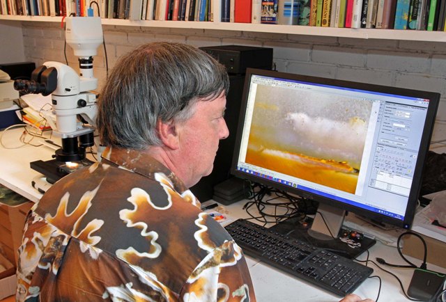
Prof Robert Weston, wearing a shirt printed with one of his ammonite fossil photographs, examines the agate crystal on the screen of the linked PC.
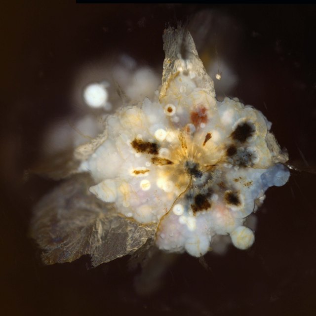
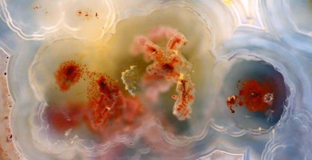
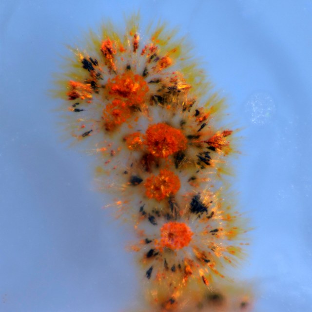
Three of Prof Weston’s photomicrographs of other agate samples. Image 5 was taken from a sub-millimeter specimen using the Z-axis capture function of the Nikon Metrology microscope.
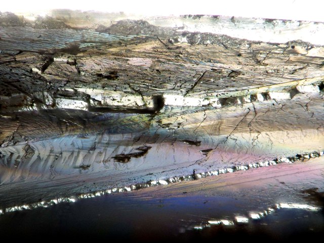
A photomicrograph of calcite, also taken using the Z-axis capture function of the Nikon Metrology microscope.
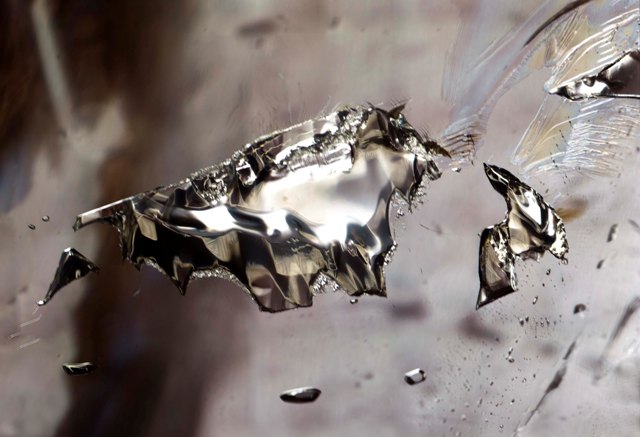
A photomicrograph of an inclusion in a quartz crystal.