Researchers at the University of Illinois have developed a computational technique, which will enable rectification of aberrations in optical tomography.
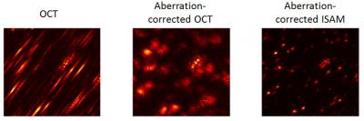 This image shows aberrations in imaging can make points appear as slashes or blurs (left frame). Computational adaptive optics developed by University of Illinois researchers can correct aberrations in high-resolution microscopy (left frames). Credit: Steven G. Adie
With this novel technology, a more affordable and higher resolution tissue imaging can be achieved. This technique has been presented by the researchers in this week’s online edition, ‘Proceedings of the National Academy of Sciences’.
High-resolution imaging has been affected by aberrations such as astigmatism or distortion, resulting in blobs- or streaks- like objects. Inaccurate or higher resolution poses threat to tissue imaging.
Adaptive optics can help rectify aberrations in imaging. Prior to entering the lens, the scattered light can be corrected using a complex system of mirrors. Promising results are being expected in cell and tissue imaging by incorporating this optics hardware to microscopes. But this hardware-based adaptive optics is highly expensive, complex, and difficult to align.
The Illinois team replaced computer software with optics hardware to correct aberrations after image capture. By partnering with Scott Carney of Beckman Institute, the Illinois research team developed a technique known as computational adaptive optics. This technique was demonstrated in gel-based phantoms impregnated with microparticles and also in rat lung tissue. The tissue sample was scanned using interferometric microscope. With the help of computer software, both collection of data and correction of images can be achieved. Computed adaptive optics is ideal for all interferometric imaging applications like optical coherence tomography. A normal desktop computer is required for these computations, and therefore can be easily adopted by hospitals and clinics.
The researchers are further planning to develop the algorithms, leading to new applications. The integration of computational adaptive optics with graphics processors will contribute to more real-time in-vivo applications in minimally invasive biopsy and other surgeries.