Aug 4 2011
Olympus has released the latest version of its virtual slide (VS) product range, the VS120. The system can rapidly scan a slide in less than two minutes, creating a digital ‘virtual slide’ that is an exact copy of the original. This is easily created and navigated using an intuitive graphical interface.
The new unit provides semi-automatic and manual focusing modes for working with challenging samples, enhanced image clarity using the new Sample Detection Mask for automatic slide scanning, and increased optimisation of user workflow. With the ability to digitise and share entire slide collections across the laboratory, classroom, or globe, the VS120 is perfect for use in research, pathology and teaching.
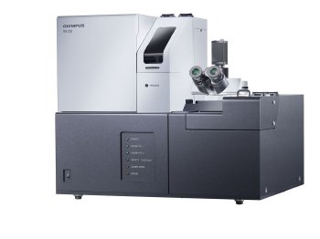 The Olympus VS120 System
The Olympus VS120 provides an array of new user benefits that simplify and enhance digital slide capture, storage and analysis. The system is extremely fast, scanning a 15 x 15mm area using a 20x objective lens with a pixel resolution of 0.345µm in around 90 seconds, while sample detail is enhanced and preserved via the high quality Olympus UIS2 optics and custom camera algorithms.
Using a threshold-based method to remove small particles, the new Sample Detection Mask improves image quality by maintaining accurate focus when using automatic settings. In addition, the Expert and Virtual Z scan modes offer semi-automatic or manual focus settings, providing more flexibility. When analysing virtual slides with multiple Z-stacks, it is possible to extract a single Z-stack of choice for further study, while the new Freehand Measurement tool can be used to generate quantifiable data from each image. To facilitate high-throughput scale-up, Olympus provides the optional VS120-L5, which can scan up to five slides automatically. For larger collections, the VS120-L100 provides ‘walk away’ scanning of up to 100 slides, while an inbuilt barcode scanner ensures that all slides are accurately tracked and catalogued.
Using the VS120, images can be captured regardless of the illumination technique, including bright-field, phase-contrast and multi-channel fluorescence via the additional fluorescence extension module. In addition, the new high resolution camera has three predefined brightfield settings that simplify and optimise the scanning of normal, dark or faintly stained samples.
The virtual slides created can be navigated digitally in real-time as if the user were controlling a microscope, without the need for an instrument or even the sample. All images are ICC profile corrected to ensure perfect colour reproduction, and the system is compatible with standard and double sized slides for maximum accuracy and flexibility. Virtual microscopy enables the information from one slide to be shared simultaneously across the world, via the simple upload interface provided by the secure Net Image Server SQL networking system. This makes the VS120 perfect for teleconsultation during disease diagnosis, global research collaboration, the archiving of slide materials and for sharing data with the classroom when teaching.