Non-invasive medical procedures, such as optical coherence tomography (OCT) imaging, are paramount in delicate organ disease management. Proper imaging eliminates invasive procedures, particularly during diagnosis. OCT is a commonly used non-invasive technique for eye care. This article explores the progress of OCT and its significance in the field of ophthalmology.

Image Credit: Chaikom/Shutterstock.com
OCT Imaging and Its Working Principle
Since its discovery in 1991, optical coherence tomography (OCT) has garnered popularity as a robust imaging modality in vitreo-retinal practice because of its non-invasive imaging ability, fast acquisition time, and high resolution. Owing to its ability to provide three-dimensional (3D) imaging of the retina and choroid, OCT has substantially contributed to the understanding of chorioretinal architecture in diverse ocular diseases.
OCT allows an ophthalmologist to view each of the retina’s distinctive layers, helping the ophthalmologist to map and measure the thickness of each layer. This measurement aids in diagnosing retinal diseases, such as glaucoma, age-related macular degeneration (AMD), and diabetic eye disease.
The underlying principle of OCT implementation is low-coherence interferometry, which is a non-contact optical sensing technology. In an OCT instrument, the sample surface is directed with low-coherence light via an optical probe and sends the reflected light signals to an interferometer via an optical fiber for interpretation.
The reflected light is analyzed, and the delay is measured to obtain the depth at which the reflection has occurred. Here, reference materials are used because delays in reflected light cannot be directly measured. The interferometer splits the reflected light into two paths and combines them back at the interferometer output.
Evolution of OCT
OCT has undergone several technical modifications, burgeoning, and upgrades. Recent advances in OCT include time-domain OCT (TD-OCT), spectral domain OCT (SD-OCT), and swept-source OCT (SS-OCT), which substantially improve the image resolution up to 5 µm.
A higher-wavelength laser with deep ocular penetration, possibly using SS-OCT, facilitates a clinician to visualize choroid-related issues, such as those involving Haller’s layer, choriocapillaris, Sattler’s layer, choroidoscleral interface, and scleral tissue, with high accuracy. SS-OCT also helps clinicians in eye-tracking and scanning features to observe changes at the pathology site.
While initial observations of the eye using OCT protocols were limited to the 6 × 6 mm macular area, introducing wide-field OCT has enhanced the coverage area, allowing the clinical utility to examine retinal degeneration, peripheral retinal ischemia, and peripheral choroidal lesions, providing additional insights into the peripheral retina.
Realizing OCTs with an increased field view and reduced acquisition time was achieved by enhancing the A-scan acquisition rate in wield-field OCT (>100,000/s) compared to the previous generation of TD-OCT (approximately 400/s). Recently, upgraded OCTs have used cutting-edge software and provided a field view of up to 20 mm.
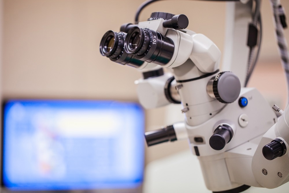
Integrating OCT imaging with surgical microscopes has added value to intraoperative anatomical assessment, which is imperative in macular surgery. Consequently, surgeons can predict surgical success rates by assessing anatomical details during intraoperative procedures.
Although the home-based and handheld versions of OCT are new additions, their low image resolution restricts their application. The non-invasive OCT technique is used in the retina, uveitis, and glaucoma clinics to quantitatively analyze the layer thickness of the retinal nerve fiber and cornea and to assess the depth of the anterior chamber.
Deep Learning (DL), Artificial Intelligence (AI), and OCT
OCT image analysis plays a crucial role in retinal disease diagnosis because the disease prediction of each eye layer is specific to a pathology. While conventional OCT image analysis is labor-intensive and time-consuming, analysis using DL and AI can drastically reduce the time and labor required to screen OCT images.
Integrating sophisticated AI and DL methods, such as support vector machine, random forest classifier, and convolutional neural network (CNN) classifier, with OCT, enhanced the resolution of OCT images.
Significant research on AI and DL-integrated OCT procedures has proved that AI is a supplemental tool to empower digital medicine but not a replacement for healthcare providers. The ability of AI to automate repetitive tasks frees physicians, creates room for more unique human tasks, and allows physicians to dedicate their time to guiding complex decisions in providing patient-centric services.
Recent Studies
A study conducted at the Hospital of Harbin Medical University investigated the potential of generative pre-trained transformers-4 (GPT-4) in recognizing retinal diseases using OCT images acquired from 80 patients undergoing treatment. Disease diagnoses were established based on previous cases, and images were uploaded to GPT-4 for diagnostic analysis.
The results revealed a consistent GTP-4 diagnostic accuracy of 26% in four distinct experiments, demonstrating a significant disparity in the results compared with the diagnostic accuracy when assessed by a retinal disease specialist (93.75%). This study revealed the limited feasibility of GPT-4 for textual information and simple images and confirmed that GPT-4 cannot substitute for professional physicians. This study was published in the journal BMC Ophthalmology.
Another study published in Advances in Ophthalmology Practice and Research evaluated choroidal thickness and retinal nerve fiber layer (RNFL) thickness in Chinese pregnant women in different trimesters by employing enhanced depth imaging OCT (EDI-OCT).
The results revealed significantly thicker subfoveal, temporal, and nasal macular choroidal parts in second-trimester pregnant women than in the first and third trimesters and control groups. The study highlighted an increased peripapillary choroidal (PPCT) thickness in second-trimester pregnant women compared with the control group at the global, temporal, temporal inferior, and nasal positions. RNFL thickness was also significantly higher in pregnant women in the nasal superior and nasal inferior quadrants.
Conclusion
Overall, OCT imaging has revolutionized ophthalmology by enhancing the quality of diagnosis and treatment monitoring. Since its inception in 1991, OCT technology has rapidly evolved, providing a higher resolution, quicker acquisition times, and deeper imaging capabilities. These developments have significantly improved our understanding of retinal and ocular diseases. The recent integration of DL and AI technologies with OCT procedures has reduced the time and labor required to assess OCT images.
Continuous enhancements in OCT technology can further refine imaging precision and broaden its clinical applications in the coming years. With faster scans and increased accuracy, OCT remains a cornerstone in ophthalmology, empowering clinicians with invaluable insights into delivering enhanced patient care.
More from AZoOptics: New X-Ray Technique Allows Effective Imaging of Living Organisms
References and Further Reading
Singh, S. R., Chhablani, J. (2022). Optical coherence tomography imaging: advances in ophthalmology. Journal of Clinical Medicine, 11(10), 2858. https://doi.org/10.3390/jcm11102858
Yu, H et al. (2023). Applications of GPT-4 for Accurate Diagnosis of Retinal Diseases Through Optical Coherence Tomography Image Recognition. BMC Ophthalmology. https://doi.org/10.21203/rs.3.rs-3644163/v1
Wu, H., Lin, H., Ruan, M., Fang, H., Dong, N., Wang, T., Zhao, J. (2023). Evaluation of choroidal thickness and retinal nerve fiber layer thickness in Chinese pregnant women and healthy non-pregnant women. Advances in Ophthalmology Practice and Research. https://doi.org/10.1016/j.aopr.2023.12.001
Wawer Matos, P. A., Reimer, R. P., Rokohl, A. C., Caldeira, L., Heindl, L. M., & Große Hokamp, N. (2023, April). Artificial intelligence in ophthalmology–status quo and future perspectives. Seminars in Ophthalmology, 38(3), 226-237. https://doi.org/10.1080/08820538.2021.1901123
Disclaimer: The views expressed here are those of the author expressed in their private capacity and do not necessarily represent the views of AZoM.com Limited T/A AZoNetwork the owner and operator of this website. This disclaimer forms part of the Terms and conditions of use of this website.