Gastrointestinal tract or gut imaging and analysis are valuable for clinical and research studies of gastrointestinal disorders. Previously, conventional radiology techniques have been dominant; however, advancements in non-invasive modalities, such as combined x-ray and photoacoustic imaging, have transformed gastrointestinal research.
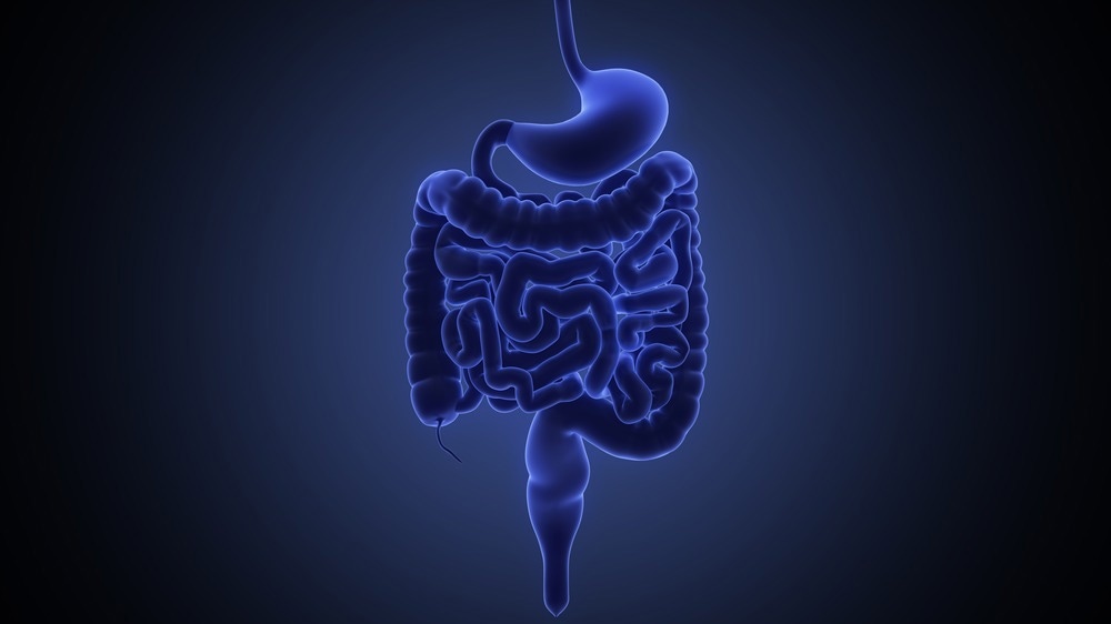
Image Credit: u3d/Shutterstock.com
Gastroenterology is a field of medical science that studies the gut or gastrointestinal system and its disorders. The gastrointestinal tract or the gut includes the mouth, esophagus, throat, stomach, large intestine, small intestine, liver, appendix, and pancreas.
Gastroenterology disorders such as inflammatory bowel disease, peptic ulcer disease, gastroesophageal reflux disease, and colorectal cancer are prevalent and serious health issues worldwide.
A physical examination and complete medical history are required to identify these disorders. However, some patients could require more comprehensive diagnostic testing, such as endoscopic procedures and imaging.
Limitations of Traditional Diagnostic Techniques
Diagnosing gastrointestinal disorders has traditionally involved invasive procedures such as biopsy, endoscopy, and colonoscopy. However, these techniques can only monitor minor inflammatory and metabolic tissue changes and detect small tumors. In addition, they have an increased risk of complications and unfavorable outcomes due to invasive procedures or contrast agents.
As a result, non-invasive diagnostic techniques such as photoacoustic imaging, computed tomography, x-rays, and magnetic resonance imaging have been introduced as alternatives to provide a more accurate and reliable analysis of the gut.
X-Ray and Photoacoustic Imaging for Gut Analysis
X-Ray Imaging
X-ray imaging is widely used for various operations and examinations. They are non-invasive and painless, making them useful for diagnosing and monitoring gastrointestinal diseases.
Barium is a contrast agent used in the x-ray diagnosis of the gastrointestinal tract. It is a white, chalky powder that is mixed with water for ingestion. When ingested, barium coats the inside of the gut, allowing the inside wall lining, contour, shape, size, and patency to be visible on X-ray for diagnosis.
The gut is exposed to a continuous X-ray beam, which is then transferred to monitor so that any organ can be diagnosed in great detail.
Photoacoustic Imaging
Photoacoustic imaging (PAI) is a non-invasive imaging technology based on the photoacoustic phenomenon, which involves the conversion of light to sound energy.
It is a simple and cost-effective alternative to other techniques such as magnetic resonance imaging, X-ray imaging, and positron emission tomography. Unlike these methods, photoacoustic imaging does not have ionizing radiation, which can adversely affect biological tissues.
Chromophores' absorption of a short-pulsed laser in tissue causes the photoacoustic effect. The absorbed laser (light energy) is transformed into thermal energy, causing the surrounding tissues to undergo thermoelastic expansion, resulting in a volumetric change.
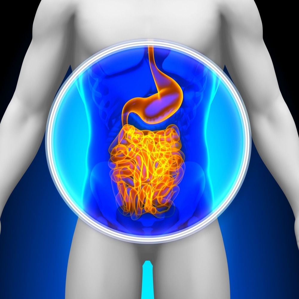
Image Credit: decade3d - anatomy online/Shutterstock.com
These volumetric changes produce vibrations that travel in the form of acoustic waves (sound energy), which are detected and processed by external ultrasonic transducers for image generation.
Combined Photoacoustic and X-Ray Imaging
Multimodal imaging is gaining interest in biomedical research as it can provide functional and anatomical information on biological tissues.
X-ray computed tomography is frequently used for cancer detection, while photoacoustic imaging has been employed to study blood vessels in mouse tumor models. These two imaging modalities can work with the same contrast agents to provide complementary information.
X-ray imaging takes advantage of the high attenuation coefficient of contrast agents. In contrast, photoacoustic imaging exploits the high sensitivity of the absorption of contrast agents in the near-infrared (NIR) region.
Combining these techniques will enable the high spatial resolution diagnosis of deep lesions.
Case Study: X-Ray and Photoacoustic Contrast Imaging of The Gut
A recent study published in the Journal of Biomedical Optics demonstrates the potential of combining photoacoustic imaging and X-ray for gut analysis with a dual-modality contrast agent. The contrast agent was a mixture of barium sulfate and near-infrared-absorbing pigments.
The mixture had a higher X-ray attenuation coefficient than barium sulfate, which enhanced the visibility of the gut organs on X-ray images. The pigment also generated a robust photoacoustic signal, allowing for more precise imaging of the gut.
In-vivo imaging of mice revealed similar patterns of contrast agent localization in photoacoustic and X-ray imaging modalities. This demonstrates that combining these two imaging modalities generates complementary insights into functional imaging of the gut, enabling the simultaneous acquisition of functional and structural data.
Concluding Remarks and Future Outlooks
A rapid and accurate gut analysis is crucial for effective treatment and improving gastrointestinal disease progression. Unfortunately, traditional imaging techniques have limitations in accurately detecting diseases, but combining photoacoustic and X-ray imaging can overcome these challenges.
This combination provides the benefits of optical and ultrasound imaging, allowing for deep tissue resolution, high penetration depth, and visualization of tissue distribution of molecules.
Although preclinical studies have shown promising results in improving clinical imaging for gastroenterology, technical challenges must be addressed before wider adoption of this technique can be anticipated. With continued improvements, however, this technique will likely have more applications in gastroenterology.
More from AZoM: Spectroscopic and Microscopic Analyses of Roman Mosaic Glass Tesserae
References and Further Reading
Kilian, H. I., Zhang, H., Bhurwani, M. M. S., Nilam, A. M., Seong, D., Jeon, M., ... & Lovell, J. F. (2023). Barium sulfate and pigment admixture for photoacoustic and x-ray contrast imaging of the gut. Journal of Biomedical Optics, 28(8), 082803. https://doi.org/10.1117/1.JBO.28.8.082803
Huang, G., Yang, S., Yuan, Y., & Xing, D. (2011). Combining x-ray and photoacoustics for in vivo tumor imaging with gold nanorods. Applied Physics Letters, 99(12), 123701. https://doi.org/10.1063/1.3643033
Bhushan, S., Anandasabapathy, S., & Petrova, E. (2020). Photoacoustic Imaging in Gastroenterology: Advances and Needs. Photoacoustic Imaging - Principles, Advances and Applications. Intechopen. https://doi.org/10.5772/intechopen.86051
Johns Hopkins Medicine. (2023). Barium X-Rays (Upper and Lower GI). [Online]. Johns Hopkins Medicine. https://www.hopkinsmedicine.org/health/conditions-and-diseases/barium-xrays-upper-and-lower-gi (Accessed on 21 February 2023)
Disclaimer: The views expressed here are those of the author expressed in their private capacity and do not necessarily represent the views of AZoM.com Limited T/A AZoNetwork the owner and operator of this website. This disclaimer forms part of the Terms and conditions of use of this website.