Photoacoustic imaging has revolutionized life sciences by providing high-speed imaging at cellular and subcellular level resolution and, therefore, has become an indispensable non-invasive tool for biomedical research.
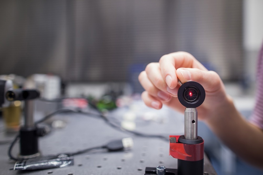
Image Credit: l i g h t p o e t/Shutterstock.com
What is Photoacoustic Imaging, and How Does it Work?
Photoacoustic imaging is a bioimaging hybrid technology that combines the benefits of ultrasound technology and optical microscopy. It exposes the tissue to pulsed laser light, causing the optical absorbers, such as hemoglobin, to expand thermally and generate an acoustic pressure wave. This wave is then detected and processed using an ultrasound transducer.
Photoacoustic imaging generates high ultrasonic and high-resolution optical contrast images. This characteristic sets it apart from other imaging methods, making it especially useful when obtaining detailed anatomical and functional images is necessary, for example, to understand key processes behind developing different diseases.
Origins and Evolution of Photoacoustic Imaging
In 1880, Alexander Graham Bell first explained the photoacoustic effect as the conversion of optical (light) energy to sound energy or waves. However, hardly any progress was achieved until the laser's invention enabled enhanced signal production. Soon after, photoacoustics became more widely used, initially for gas spectroscopy and later in life sciences.
During the last two decades, photoacoustic imaging has rapidly progressed from a theoretical concept to a widely applied tomographic method in biomedical applications.
How is Photoacoustic Imaging used in the field of Life Sciences?
Brain Imaging
One of the most significant biological applications of photoacoustic imaging is in vivo functional and structural brain imaging. Photoacoustic imaging offers higher molecular and resolution imaging capacity when coupled with biomarkers than other functional imaging technologies.
Unfortunately, strong light scattering by the brain tissue and skull poses a major challenge for the technology. Currently, photoacoustic brain imaging is still in its preclinical stage and is primarily being tested on rodents due to their thinner skulls.
However, advancements such as Nie et al.'s development of a photon recycler, which increases light transmittance through the skull, have improved the photoacoustic imaging signal-to-noise ratio and made it feasible to study functional activity in the human cerebral cortex.
Vascular Imaging
The World Health Organization (WHO) has identified cardiovascular disease as one of the leading causes of death for the past decade. Lipid-rich atherosclerotic plaques have been identified as a cause of cardiovascular disease, and photoacoustic imaging's high optical absorption at 1.7μm and 1150–1250 nm are ideal for distinguishing lesions from healthy tissues.
Blood Oxygenation and Flow
In cancer research, dermatology, and plastic surgery, measuring blood oxygenation levels in the skin is an essential physiological metric. Near-infrared spectroscopy is the most extensively used in vivo method for measuring blood oxygenation; however, it has a low spatial resolution. On the other hand, photoacoustic imaging provides higher resolution in biological tissues, making it a suitable alternative.
A study published in Proceedings of the National Academy of Sciences demonstrated that photoacoustic imaging could explain the relationship between oxygen delivery and neural activity in response to various physiological conditions, providing valuable insight into how the brain is powered at the cellular level.
Breast Imaging
Breast cancer is the leading cause of cancer-related deaths in women. However, current cancer screening techniques have limited specificity, low positive predictive value, and cause extreme discomfort.
Photoacoustic imaging has great potential to improve the diagnostic imaging of the breast. The breast tissue is located on the body's surface and falls within the photoacoustic imaging's resolution. In addition, healthy breast tissue has low ultrasonic scattering and optical absorption, making photoacoustic imaging highly effective.
Moreover, angiogenesis plays a significant role in diagnosing and predicting breast cancer. Again, photoacoustic imaging is well-suited for visualizing this because the increased hemoglobin and blood flow at tumor sites create strong contrast in photoacoustic imaging.
Recent Research and Development
Imaging-Based Machine Learning Model Improves Ovarian Lesion Detection
Yun Zou and his team from Washington University designed a new machine learning method that combines ultrasound and photoacoustic imaging neural networks to diagnose ovarian lesions.
The team enhanced the accuracy of ultrasound diagnosis by incorporating the blood oxygenation saturation and total hemoglobin concentration (indicators of cancerous ovarian tissue) obtained from photoacoustic imaging.
Our results showed that the ultrasound-enhanced photoacoustic imaging fusion model reconstructed the target's total hemoglobin and blood oxygen saturation maps more accurately than other methods and provided an improved diagnosis of ovarian cancers from benign lesions
Yun Zou, Study Main Author and Doctoral Student, Washington University
3D Bimodal Photoacoustic Ultrasound Imaging for Peripheral Vascular Disease Diagnosis
Conventional techniques, such as the ankle-brachial index test, can detect anomalies in major arteries, but they struggle to diagnose the numerous thin peripheral blood vessels. In addition, they can potentially cause discomfort or adverse effects because a contrast agent has to be administered to the patient.
To address these limitations, researchers from Pohang University of Science & Technology have developed a novel 3D bimodal photoacoustic imaging technique by combining ultrasound and photoacoustic imaging to produce 3D images of the blood vessels without using a contrast agent.
This new technique can potentially diagnose peripheral vascular diseases by offering crucial functional diagnostic values such as blood oxygen saturation without side effects.
Using Photoacoustic Imaging to Reveal Rapid Brain Activity
Duke University biomedical engineers have designed the fastest photoacoustic imaging technology to scan and monitor the oxygen levels and blood flow inside a rodent's brain in real-time with high resolution. This tool allows for the simultaneous visualization of individual vessels and the entire brain.
The improvement was achieved through machine learning algorithms and hardware enhancement. The researchers used a polygon scanning system to deliver more laser bursts to a larger area and a new scanning method to run the ultrasound sensor and laser scanner simultaneously. These modifications doubled its imaging speed and made it the fastest photoacoustic imaging technology available.
This innovation removes long-standing resolution and speed constraints in brain imaging technology and will reveal new insights into neurovascular diseases such as dementia and stroke.
Future Outlooks of Photoacoustic Imaging
The development of photoacoustic imaging systems has provided new opportunities for disease monitoring, treatment, and diagnosis in life sciences. By using advanced computational analyses, the evaluation of the large amounts of data generated by these systems can be improved beyond just visual interpretation in clinical diagnostics.
Even though current systems still have limitations, such as time-consuming spectral un-mixing and image reconstruction algorithms. Improving these processes can lead to faster processing times and better image interpretability, enhancing the overall usability of photoacoustic imaging systems.
More from AZoOptics: What is Photothermal Spectroscopy?
References and Further Readings
Neprokin, A., Broadway, C., Myllylä, T., Bykov, A., & Meglinski, I. (2022). Photoacoustic imaging in biomedicine and life sciences. Life, 12(4), 588. https://doi.org/10.3390/life12040588
Steinberg, I., Huland, D. M., Vermesh, O., Frostig, H. E., Tummers, W. S., & Gambhir, S. S. (2019). Photoacoustic clinical imaging. Photoacoustics, 14, 77-98. https://doi.org/10.1016/j.pacs.2019.05.001
Zou, Y., Amidi, E., Luo, H., & Zhu, Q. (2022). Ultrasound-enhanced Unet model for quantitative photoacoustic tomography of ovarian lesions. Photoacoustics, 28, 100420. https://doi.org/10.1016/j.pacs.2022.100420
Choi, W., Park, E. Y., Jeon, S., Yang, Y., Park, B., Ahn, J., ... & Kim, C. (2022). Three-dimensional multistructural quantitative photoacoustic and US imaging of human feet in vivo. Radiology, 303(2), 467-473. https://doi.org/10.1148/radiol.211029
Zhu, X., Huang, Q., DiSpirito, A., Vu, T., Rong, Q., Peng, X., ... & Yao, J. (2022). Real-time whole-brain imaging of hemodynamics and oxygenation at micro-vessel resolution with ultrafast wide-field photoacoustic microscopy. Light: Science & Applications, 11(1), 138. https://doi.org/10.1038/s41377-022-00836-2
Nie, L., Cai, X., Maslov, K., Garcia-Uribe, A., Anastasio, M. A., & Wang, L. V. (2012). Photoacoustic tomography through a whole adult human skull with a photon recycler. Journal of biomedical optics, 17(11), 110506-110506. https://doi.org/10.1117/1.JBO.17.11.110506
Wang, L., Maslov, K., & Wang, L. V. (2013). Single-cell label-free photoacoustic flowoxigraphy in vivo. Proceedings of the National Academy of Sciences, 110(15), 5759-5764. https://doi.org/10.1073/pnas.1215578110
Washington University in St. Louis. (2022). Machine learning model builds on imaging methods to better detect ovarian lesions. ScienceDaily. Retrieved February 2, 2023 from www.sciencedaily.com/releases/2022/11/221129134412.htm
Disclaimer: The views expressed here are those of the author expressed in their private capacity and do not necessarily represent the views of AZoM.com Limited T/A AZoNetwork the owner and operator of this website. This disclaimer forms part of the Terms and conditions of use of this website.