Image Credit: Micha Weber/Shutterstock.com
Conventional fluorescence lifetime imaging microscopy (FLIM) relies on point-to-point scanning of a tightly focused illumination beam across the sample to form an image, impeding rapid imaging. To overcome this limitation, researchers from Japan and the USA have developed scan-less wide-field imaging methods that can image fluorescence lifetime at unprecedented speeds and capture ultrafast transient phenomena relevant to biomedical research and life sciences.
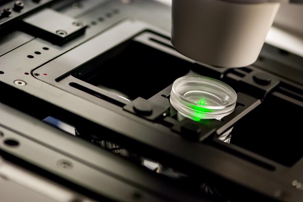
FLIM is an imaging technique that creates spatially resolved images of a fluorophore (fluorescent molecule used as a marker) lifetime. The fluorescence lifetime is an intrinsic property of the fluorophore, which describes, on average, how rapidly the fluorophore's excited state decays.
FLIM Expands the Functionality of the Conventional Fluorescence Microscopy
Being a non-invasive, non-destructive, and sensitive technique, FLIM has become an analytical tool of choice for biomedical research and material characterization. In particular, FLIM has seen an ever-increasing use in high-throughput, high-content drug screening, clinical diagnostics, and for the study of innovative materials and biochemical reactions.
FLIM's main advantage over the intensity-based fluorescence microscopy is the ability to discriminate spectrally overlapping fluorophores by time-resolved measurements of the fluorophore's lifetime.
The acquisition rate of the FLIM data is a complex function of many factors. These include, in the first place, the scanning rate of the illumination beam across the sample, the number of emitted photons (without causing photodamage to the specimen), the accuracy of fluorescence decay parameters determination, and the required spatial resolution.
FLIM's slow data acquisition rate (typically several minutes per image) is a significant downside, limiting the technique's use when observing fast transient processes, such as small moving micro-organisms, the motion of cellular organelles, trafficking of vesicles and proteins, and other fast biophysical phenomena.
Dynamic Biological Processes Require Faster Imaging Rates
There is a significant drive to minimize FLIM data acquisition time that will permit increased temporal sampling of dynamic events or high-throughput screening applications.
Fluorescence lifetime can be measured either by time-correlated single-photon counting (TC-SPC), where single photons arrival times are correlated to a reference pulsed excitation, or by a phase measurement (PM) method, where the fluorescent lifetime is derived from the phase shift of the fluorescence signal relative to a sinusoidally-modulated excitation light beam.
Since both methods are based on point-to-point measurements, mechanical scanning of the microscope's focal point is necessary to obtain an image. Such an optomechanical scanning system often limits the overall imaging rate of a FLIM system.
A recent breakthrough development by a research group from the Institute of Post-LED Photonics at Tokushima University in Japan, led by Prof. Takeshi Yasui, promises to revolutionize fluorescence lifetime imaging by enabling massively parallel rapid quantitative FLIM without the need for mechanical sample scanning.
Optical Frequency Combs Replace Multiple Individual Light Sources
Prof. Yasui's team combined PM lifetime measurements with parallelized frequency-multiplexed excitation of the sample. By employing an optical frequency comb (OFC) that converts a continuous light spectrum into a stream of numerous co-existing beams with equally spaced wavelengths, the researchers created many separate light beams.
Superimposing and tuning the two optical combs' discrete spectra resulted in a dual-comb optical beating (interference between the mixed frequencies). They generated an ultradense array of 44,400 frequency-multiplexed individual light beams illuminating the entire sample surface simultaneously.
The sample re-emits part of excitation light while maintaining the frequency-position encoding. The encoded fluorescence signal is collected and imaged onto a high-speed single-point photodetector.
Simultaneous Fluorescence Lifetime Measurements across the Entire Sample Surface
The direct correspondence between the dual-comb frequencies and the beams' spatial position (meaning that each beam possesses a unique modulation frequency) allowed the scientists to decode each beam's amplitude and phase shift (representing a single image pixel). They created a 2D image representing the fluorescence intensity and lifetime across the specimen surface.
According to Prof. Yasui, the new dual-comb FLIM technique offers far superior imaging speed while maintaining high spatial resolution. Because the dial-comb FLIM does not require scanning, it permits a one-shot fluorescence lifetime measurement over the entire sample surface.
Using a state-of-the-art low-noise CMOS (complementary metal-oxide-semiconductor) camera, the researchers have achieved an equivalent imaging rate of 45 frames per second. However, according to Prof. Yasui and his colleagues, the technique can perform at frame rates of up to 1000 frames per second.
Scan-Less Full-Field FLIM for Parallelized Diagnostics
The research team explores a further increase of the image resolution through pixel interleaving, available via the dual-comb beat frequency tunability. This enables simultaneous imaging of multiple fluorophores at different wavelengths.
Besides providing a better understanding of many biological processes, the new approach is applicable for simultaneous imaging of large arrays containing multiple samples for antigen testing, a method already used for COVID-19 diagnosis.
References and Further Reading
T. Mizuno, et al. (2021) Full-field fluorescence lifetime dual-comb microscopy using spectral mapping and frequency multiplexing of dual-comb optical beats. Science Advances, 7, eabd2102. Available at: https://doi.org/10.1126/sciadv.abd2102
C. Poudel, et al. (2020) High-throughput, multi-parametric, and correlative fluorescence lifetime imaging. Methods Appl. Fluoresc., 8, 024005. Available at: https://doi.org/10.1088/2050-6120/ab7364
Tokushima University (2021) Comb of a Lifetime: A New Method for Fluorescence Microscopy [Online] www.pled.tokushima-u.ac.jp Available at: https://www.pled.tokushima-u.ac.jp/pressrelease-6/2298 (Accessed on 7 February 2021).
Photonics Media (2021) Technique Increases Effectiveness of Fluorescence Lifetime Microscopy. [Online] www.photonics.com Available at: https://www.photonics.com/Articles/Technique_Increases_Effectiveness_of_Fluorescence/a66571 (Accessed on 7 February 2021).
X. Liu, et al. (2019) Fast fluorescence lifetime imaging techniques: A review on challenge and development. J. Innov. Opt. Heal. Sci., 12, 1930003. Available at: https://doi.org/10.1142/S1793545819300039
Disclaimer: The views expressed here are those of the author expressed in their private capacity and do not necessarily represent the views of AZoM.com Limited T/A AZoNetwork the owner and operator of this website. This disclaimer forms part of the Terms and conditions of use of this website.