Image Credit: PolakPhoto/Shutterstock.com
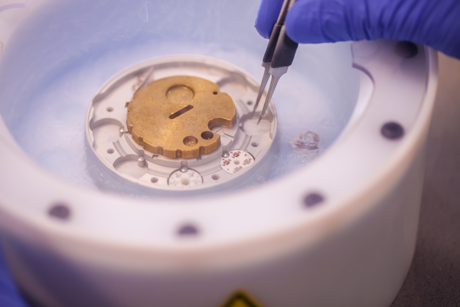
Cryo-electron microscopy (EM) has recently emerged as one of the main techniques used to study the structural biology of many macromolecular complexes at near-atomic resolution. More recently, this high-resolution technique has been incorporated into the drug discovery process for a number of different purposes.
What is Cryo-EM?
Since its original discovery in 1945, EM has remained an invaluable tool for the analysis, evaluation, and discovery of biological specimens. Despite its numerous advantages, EM techniques have often been associated with limitations as a result of hardware constraints and the inherent nature of biological samples that can cause difficulties during EM sample preparation.
Because the electron scattering that occurs within an EM requires the column to be under a vacuum, biological specimens, which are often naturally present in aqueous environments, must undergo length drying and processing procedures prior to imaging. In an effort to overcome some of these limitations, researchers in 1975 began experimenting with how traditional EM techniques could be used to analyze frozen samples.
Recent Developments in Cryo-EM
Until recently, nuclear resonance spectroscopy (NMR) and X-ray crystallography were considered the only techniques capable of providing detailed information on the structural characteristics and molecular interactions of biological samples. As technology has advanced, the development of direct electron detectors has played a critical role in the recent revolution of cryo-EM applications in biological sciences.
These technologies have subsequently allowed cryo-EM to be used in the analysis of non-crystalline single-particle specimens at near-atomic resolution. Simultaneously, researchers have been able to utilize cryo-EM for the discovery of multiple functionally relevant conformations that may be present within a single mixture.
Cryo-EM vs. X-Ray Crystallography and NMR
While the use of both NMR and X-ray crystallography have allowed for pharmaceutical companies to obtain structural information on a wide range of target molecules, there remains little to no structural information on a considerable number of targets.
This lack of information is largely attributed to the molecular size, complexity, flexibility, and the difficulties associated with the production and purification of these other targets, all of which make it impossible for researchers to obtain structure information from NMR and/or crystallography. Furthermore, structural information obtained by X-ray crystallography typically only represents a single conformation of the target protein, which may not reflect its conformation when present at the cellular level.
Comparatively, cryo-EM is capable of accessing larger and even more complex biological systems as compared to both X-ray crystallography and NMR. Furthermore, cryo-EM has been found to allow users to analyze proteins and characterize multiple conformational and/or compositional states of molecules from a single sample, thereby demonstrating its superiority to traditional structural techniques.
Importance of Structural Biology in Drug Development
Structure-guided drug design plays a central role in the drug development process and can take several years to achieve before a new drug candidate is obtained. This specific aspect of the drug design process requires structural, computational, and medicinal chemists, biologists, and pharmacologists to be involved. Although traditional techniques like X-ray crystallography and NMR can be used to answer relevant biological questions despite being at a lower-resolution, the specific requirements of the structure-guided drug design requirement eliminate these techniques from being useful.
At the earliest stages of drug development, which is otherwise referred to as the target identification stage, cryo-EM can be used to provide the user with information on how a specific target can be activated and/or modulated. From here, researchers can incorporate this information into other types of early drug development assays, including in silico screening, docking, and design to determine whether any pharmacophores or optimal binding vectors should be incorporated into the new drug.
As a given drug moves past target and hit identification stages, structural biology becomes an even more essential aspect of the drug development process. From here, researchers are expected to provide structures of a given target molecule when bound to pharmacophores. Furthermore, any simple or large chemical change to a given drug will require repeated structural investigation to investigate whether the given modification has caused any major pharmacological effects to arise.
Cryo-EM in Drug Development
It is evident that early drug development processes require high-resolution imaging techniques that are capable of detecting subtle structural changes in any new drug target. The advantages associated with cryo-EM have led numerous pharmaceutical and biotechnology companies around the world to embrace this technique for the structural determination of novel drug targets.
One example of how cryo-EM has revolutionized the way in which drug development is carried out, can be found in a recent study on mefloquine (MFQ), which is a common antimalaria medication. When the group of researchers studying MFQ turned to cryo-EM over conventional structural analysis techniques, they found that this target binds to Plasmodium falciparum 80S ribosome, which was not previously known. As a result, the researchers utilized this information to modify the structure of MFQ, create several derivatives, and ultimately increase its potency for future malaria treatments.
Future of Cryo-EM
Because the reinvigoration of cryo-EM only recently began in 2013, not all industrial and academic scientists have taken advantage of this technology for their structural analysis of new drug targets. Nevertheless, the constantly-improving software and hardware of this technology supports the promising outlook of cryo-EM-driven drug development investigation.
References and Further Reading
Egelman, E. H. (2016) The Current Revolution in Cryo-EM. Biophysical Journal 110(5); 1008-1012. DOI: 10.1016/j.bpj.2016.02.001.
Subramaniam, S., Earl, L. A., Falconieri, V., Milne, J. L. S., & Egelman, E. H. (2016) Resolution advances in cryo-EM enable application to drug discovery. Current Opinion in Structural Biology 41; 194-202. DOI: 10.1016/j.sbi.2016.07.009.
Scapin, G., Potter, C. S., & Carragher, B. (2018) Cryo-EM for Small Molecules Discovery, Design, Understanding and Application. Cell Chemical Biology 25(11); 1
SelectScience (2020) How the pharma industry is harnessing cryo-electron microscopy in drug discovery. Available at: https://www.selectscience.net/editorial-articles/how-the-pharma-industry-is-harnessing-cryo-electron-microscopy-in-drug-discovery/?artID=50611 (Accessed on 6 March 2020).
Disclaimer: The views expressed here are those of the author expressed in their private capacity and do not necessarily represent the views of AZoM.com Limited T/A AZoNetwork the owner and operator of this website. This disclaimer forms part of the Terms and conditions of use of this website.