Cryo-electron microscopy (cryo-EM) allows for the visualization of complex biological structures at near-atomic resolution.
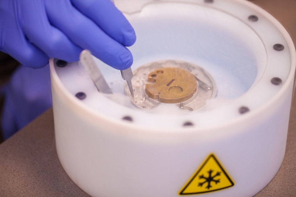
Image Credit:PolakPhoto/Shutterstock
One main objective of structural biology is to provide a mechanistic understanding of biological processes. The atomic structures of macromolecules and complexes in functional states offer the most thorough information. In addition to basic research, the pharmaceutical industry uses atomic structure data of drug targets to design and optimize therapeutic drugs.
Traditional Molecular Imaging Techniques
Various experimental techniques have been developed to study molecular structures in more detail. X-Ray crystallography has historically been used to examine biological structures, but this method requires the molecule to crystallize, which can be challenging or impossible for many proteins.1
Nuclear Magnetic Resonance (NMR) provides high-definition images of biomolecules but is limited to very small proteins or specific regions of molecules. Optical microscopes have also played a significant role, but diffraction limits restrict their resolution.
Transmission electron microscopes (TEMs) use an electron beam to view biomolecule structures at the atomic scale. The resolution achievable with a TEM is significantly higher than that of an optical microscope because the wavelength of the radiation source directly influences the resolution of a microscope. However, certain materials, particularly biomolecules, cannot withstand the intense electron beam and vacuum conditions of a TEM.
Cryo-Electron Microscopy: Techniques and Methods
The shortcomings of traditional electron microscopy led to the invention of cryo-EM. Cryo-EM captures the three-dimensional (3D) structure of biological molecules and complexes with near-atomic resolution.
This method traps the material in vitreous ice by quickly freezing it to temperatures below -150 °C.2 The sample is then imaged from various perspectives using an electron microscope, producing a series of two-dimensional (2D) projections. These projections are used to reconstruct a 3D model using advanced computational processes.
Developed through the pioneering work of scientists like Jacques Dubochet, Joachim Frank, and Richard Henderson, cryo-EM has become a powerful tool, earning the 2017 Nobel Prize in Chemistry.
The cryo-EM workflow begins with preparing a sample, usually a pure protein or complex in solution. The sample solution is submerged in a cryogenic fluid, such as liquid ethane, and quickly frozen after being arranged on a specific grid.3
The vitrified sample is then placed in the cryo-EM microscope, where it is exposed to an electron beam that creates a sequence of 2D projection images from various viewpoints. These 2D images are processed to increase the signal-to-noise ratio and correct for artifacts. Computational tools are then used to create a 3D density map representing the electron scattering intensities of the material.
Cryo-EM is used to research viral structures to develop vaccines and antivirals, understand protein interactions in biological functions, examine complex molecules involved in diseases, and design novel medications targeting specific proteins.
Types of Cryo-EM
There are several types of Cryo-EM, each applied to studying biological structures in different settings.
- Electron Crystallography: This technique is beneficial for studying tiny membrane proteins, which are crystallized in 2D arrays so that cryo-EM data may be collected and processed later.4
- Cryo-Electron Tomography (Cryo-ET): This technique views a biological sample that has been flash-frozen and thinned to the proper thickness. The material is kept near-native and hydrated. Several images are taken as the sample is tilted along an axis, and the data is combined and aligned computationally to create a 3D tomogram.5
- Single-Particle Cryo-EM: This method determines structures by computationally integrating images of numerous individual macromolecules in the same or similar conformations. It has become viable for atomic resolution structure determination at resolutions better than 4Å.
Future Outlook
Cryo-EM is increasingly used to study protein assemblies, viruses, and cells at molecular resolution. Advances in imaging technology, microscope design, and improved image processing and automation are enhancing its efficacy.
These advancements should enable the technology to determine a broad range of biological structures by increasing the speed and scope of automation and improving the attainable resolutions.
More from AZoOptics: Advancing Cancer Diagnosis with Optical Genome Mapping
References and Further Reading
- Cheng Y. (2015). Single-Particle Cryo-EM at Crystallographic Resolution. Cell. doi.org/10.1016/j.cell.2015.03.049
- Milne, JL., Borgnia, MJ., Bartesaghi, A., Tran, EE., Earl, LA., Schauder, DM., Lengyel, J., Pierson, J., Patwardhan, A., Subramaniam, S. (2014). Cryo-electron microscopy--a primer for the non-microscopist. FEBS J. doi.org/10.1111/febs.12078
- Carreras, HZ. (2023) Cryo Electron Microscopy: Principle, Strengths, Limitations and Applications. [Online] Technology Networks. Available at: https://www.technologynetworks.com/analysis/articles/cryo-electron-microscopy-principle-strengths-limitations-and-applications-377080#D2
- Uddin, YS., Schmidt-Krey, I. (2015). Chapter Seventeen - Inducing Two-Dimensional Crystallization of Membrane Proteins by Dialysis for Electron Crystallography. Methods in Enzymology. doi.org/10.1016/bs.mie.2014.12.022.
- Doerr, A. (2017). Cryo-electron tomography. Nat Methods. doi.org/10.1038/nmeth.4115
Disclaimer: The views expressed here are those of the author expressed in their private capacity and do not necessarily represent the views of AZoM.com Limited T/A AZoNetwork the owner and operator of this website. This disclaimer forms part of the Terms and conditions of use of this website.