Reviewed by Lexie CornerJul 9 2025Reviewed
Researchers have developed a prototype imaging system that may improve the ability of doctors to detect malignant tissue during endoscopic procedures. The findings were published in the Journal of Medical Imaging.
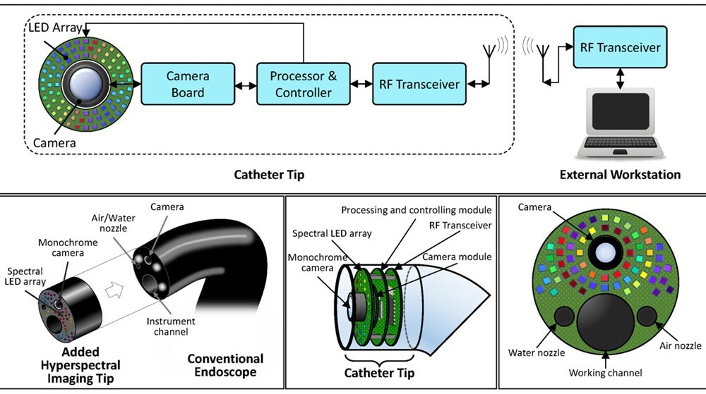 The proposed hyperspectral endoscopic imaging system includes a spectral LED array in the catheter tip. Image Credit: N. Modir et al., doi 10.1117/1.JMI.12.3.035002
Gastrointestinal cancers are among the most common types of cancer. Although endoscopy has been a standard method for screening and diagnosis for the past 20 years, it still fails to detect 8 to 11 percent of cancers. This is due to limitations in visual detection.
The new system uses a combination of light-emitting diodes (LEDs) and hyperspectral imaging to produce detailed maps of tissue properties. These features are not visible with conventional endoscopic cameras.
Traditional endoscopy captures images using broad red, green, and blue (RGB) channels. In contrast, hyperspectral imaging collects data across many narrow wavelength bands, including light outside the visible range. This allows the system to detect metabolic changes in cancerous cells, which create distinct spectral patterns.
Testing the LED Array Concept
The research team, led by Dr. Baowei Fei, Professor and Cecil H. and Ida Green Chair in Systems Biology Science at the University of Texas at Dallas Quantitative BioImaging Laboratory, developed and tested a prototype imaging system. The system uses an array of 18 LEDs that emit light at wavelengths between 405 and 910 nanometers.
As each LED illuminates the tissue one at a time, a monochrome camera captures images. This process generates a complete hyperspectral dataset.
The team evaluated the system by imaging both normal and cancerous tissue samples collected after surgery. They tested different imaging settings to assess how these conditions affected data quality. Results from their system were compared to a reference hyperspectral camera, which served as the gold standard.
Promising Results for Medical Applications
The LED-based prototype successfully captured hyperspectral signals from different tissue types. Its performance was comparable to that of the reference system. The researchers found that their method could achieve imaging speeds of over 10 hyperspectral datasets per second. This meets the real-time requirements for endoscopic procedures.
Our research shows the feasibility of employing a spectral LED array as the illumination source for high-speed and high-quality hyperspectral imaging. Our findings suggest that LED-based systems could open new possibilities for hyperspectral imaging applications.
Naeeme Modir, Study First Author and Ph.D. Candidate, University of Texas
Advantages Over Current Technology
The LED-based system offers several advantages over traditional hyperspectral endoscopy devices. Conventional systems often rely on fiber optic bundles to deliver light through the endoscope’s working channel. This setup reduces space for surgical tools or other instruments.
In contrast, the new design places the LEDs directly at the tip of the endoscope. This leaves the working channel available for medical instruments or other functions.
The system also allows real-time adjustment of each LED’s intensity based on the distance to the target tissue. This helps prevent sensor saturation caused by excessive light and reduces noise from underexposure. Additionally, because each wavelength is applied for only a short time, the design may reduce the risk of prolonged light exposure compared to systems using broad-spectrum white light.
Technical Innovation and Future Applications
The researchers used a wavelength-scanning method, where LEDs illuminate the tissue one wavelength at a time. This approach balances the need for fast imaging with the need for high spectral resolution. Unlike existing hyperspectral systems, which often trade speed for resolution or vice versa, this method maintains both.
The use of micro-LED technology makes the device suitable for medical applications. These LEDs are smaller than 400 micrometers on each side and can be mounted on a circuit board around the camera at the tip of the endoscope. This compact design allows multiple LEDs to be integrated without significantly increasing the size of the endoscope.
Another benefit is the ability to select specific spectral bands for imaging. Clinicians can choose wavelength combinations that are most effective for detecting certain cancers or tissue abnormalities. This can improve diagnostic accuracy while keeping imaging speeds high.
Path Forward
Modir added, “While the prototype demonstrates the technical feasibility of LED-based hyperspectral endoscopy, additional development will be needed before the technology reaches clinical practice. We established that this approach can produce high-quality hyperspectral data at practical imaging speeds - hopefully providing a foundation for future medical applications.”
This study represents a step toward more effective endoscopic cancer screening. LED-based hyperspectral imaging could reduce the number of missed tumors and support real-time tissue assessment over larger areas. This may help physicians make faster and more accurate decisions while minimizing unnecessary tissue removal and lab testing.
Journal Reference:
Modir, N., et al. (2025) LED-based, real-time, hyperspectral imaging device. Journal of Medical Imaging. doi.org/10.1117/1.JMI.12.3.035002.