Researchers have developed a new surgical microscope, called FiLM-Scope, that uses 48 cameras and advanced algorithms to generate precise, real-time 3D maps of the surgical field - overcoming key limitations of traditional stereoscopic imaging.
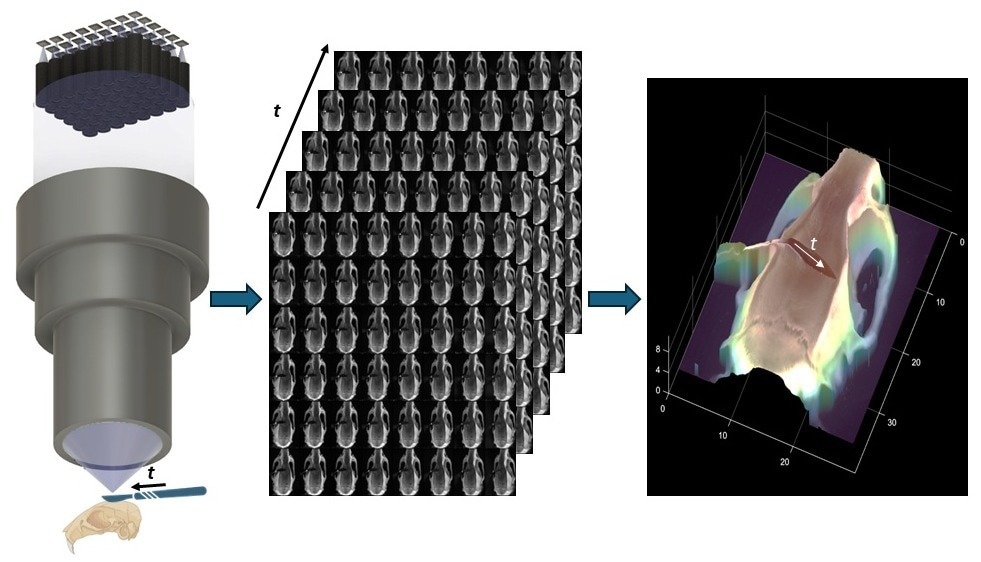 FiLM-Scope simultaneously captures 48 multiperspective images of a surgical scene. Using a custom reconstruction algorithm, these images can be converted into a dense 3D model. Image Credit: Clare B. Cook | Duke University
For more than a century, surgeons have relied on stereoscopic microscopes to gain depth perception during delicate procedures. These tools mimic human vision by showing each eye a slightly different image, enabling the brain to reconstruct a three-dimensional view - a crucial advantage when navigating fragile blood vessels or intricate brain structures. Despite modern upgrades like digital displays and video recording, today’s operating microscopes still rely on that same core principle: two images interpreted by the human brain.
But that method has its limitations. While it offers a strong intuitive sense of depth, it doesn’t allow surgeons - or surgical robots - to extract precise spatial measurements from the images. Accurately determining distances or object shapes using just two perspectives is tough, especially in complex surgical settings with uneven lighting, reflective surfaces, and obstructing tools. These challenges have slowed the development of surgical automation and real-time visual feedback systems.
Pre-operative 3D scans such as MRIs and CTs provide valuable context, but they can’t adjust to dynamic changes during surgery. Optical coherence tomography (OCT) delivers high-resolution, real-time data, but only for a small area, and its grayscale images can be hard to interpret in a fast-paced operating room.
FiLM-Scope addresses these gaps with a novel approach. The system uses a grid of 48 miniature cameras, all focused through a single, high-throughput lens. Each camera captures the surgical field from a slightly different angle, generating 48 high-resolution images (12.5 megapixels each) of the same scene. The field of view is broad - around 28 by 37 millimeters - with detail sharp enough to resolve structures as small as 22 microns. It also supports video at up to 120 frames per second.
These multiple perspectives are processed in real time by a self-supervised algorithm that constructs a detailed 3D map of the scene - no prior training data or models required. The algorithm can measure surface shapes with 11-micron accuracy across a depth range of one centimeter. Because each frame contains full-angle coverage, surgeons can digitally zoom or shift perspective without physically adjusting the microscope, making procedures smoother and more efficient.
By turning visual data into exact 3D measurements, FiLM-Scope opens up new possibilities for both manual and robotic microsurgery. Its flexible, high-resolution imaging could also benefit fields that demand ultra-precise 3D visualization, such as materials science, microfabrication, or biological research.
Journal Reference:
Cook, C., B., et al. (2025) Fourier lightfield multiview stereoscope for large field-of-view 3D imaging in microsurgical settings. Advanced Photonics Nexus. doi.org/10.1117/1.APN.4.4.046008.