Apr 29 2021
Analyzing proteins at low concentrations, particularly those in a mixture of different conformations like intrinsically disordered proteins (IDPs), can be challenging.
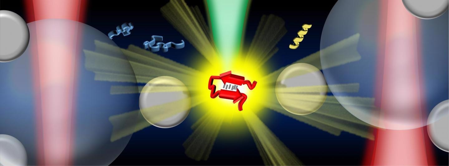
 An illustration showing the optical tweezers-controlled hotspot for the protein structural characterization by surface-enhanced Raman spectroscopy. Image Credit: Vince St. Dollente Mesias, Jinqing Huang / The Hong Kong University of Science and Technology.
A team of researchers guided by Prof. Jinqing Huang, Assistant Professor at the Department of Chemistry of The Hong Kong University of Science and Technology (HKUST), has created optical tweezers-coupled Raman spectroscopy capable of directly probing the structural properties of alpha-synuclein, an IDP closely associated with Parkinson’s disease, at the physiological concentration by centering on distinct protein molecules.
IDPs have a vital role to play in biological processes and several of them are linked with incurable neurodegenerative diseases. As a characteristic IDP, alpha-synuclein does not possess a stable 3D architecture referred to as secondary structures. It automatically undergoes a change from one secondary structure to another, which could ultimately lead to the accumulation of protein aggregates found in Parkinson’s disease pathology.
But the transient species during the conversion comprise numerous structures and are present in a low population among a dynamic equilibrium mixture. Hence, their structural features are often suppressed under the detection results from conventional measurement methods, which average the signals identified from large sample quantities and long detection time.
As part of the research, Prof. Huang and her colleagues combined surface-enhanced Raman spectroscopy (SERS) and optical tweezers into a unique platform to produce tunable and reproducible SERS improvements with single-molecule level sensitivity in aqueous settings, to depict these IDPs while preserving their inherent heterogeneity with excellent biological implication.
A hotspot can specifically be pictured and regulated by the optical tweezers to enable proteins to pass into a microfluidic flow chamber. This makes it easy to alter the measurement parameters in real time for the in situ spectroscopic classifications.
It instantly detects the structural features of the transient species of alpha-synuclein among its principal monomers at a physiological concentration of 1 μM by decreasing the ensemble averaging in time and quantity. This offers deep insights to understand the start of amyloid protein aggregation.
Therefore, this SERS platform has significant potential to expose the structural data of IDPs in the dynamic, heterogeneous, and multifaceted biological systems.
Our strategy enables the precise control of the hotspot between two trapped micrometer-size silver nanoparticle-coated silica beads to improve the SERS efficiency and reproducibility in aqueous detections. Except for the tunable SERS enhancement, the integrated optical tweezers also offer sub-nanometer spatial resolution and sub-piconewton force sensitivity to monitor light-matter interactions in the plasmonic hotspot for extra physical insight.
Jinqing Huang, Study Lead and Assistant Professor, Department of Chemistry, Hong Kong University of Science and Technology
“More importantly, our method opens a new door to characterize the transient species of IDPs in dilute solutions, which remains a significant challenge in the biophysics community,” added Prof. Huang.
Ultimately, it will be exciting to fully exploit the precise force manipulation of the integrated optical tweezers to unfold a single protein inside the controllable hotspot and resolve its structural dynamics from the endogenous molecular vibrations by the integrated Raman spectroscopy.
Jinqing Huang, Study Lead and Assistant Professor, Department of Chemistry, Hong Kong University of Science and Technology
Journal Reference:
Dai, X., et al. (2021) Optical tweezers-controlled hotspot for sensitive and reproducible surface-enhanced Raman spectroscopy characterization of native protein structures. Nature Communications. doi.org/ 10.1038/s41467-021-21543-3.