Oct 4 2017
A research carried out at Karolinska Institutet and Karolinska University Hospital indicates that an innovative microscopy method for three-dimensional investigation of tumor tissue has the ability to more precisely diagnose cancer than prevalent two-dimensional techniques. The study has been published in the Nature Biomedical Engineering journal.
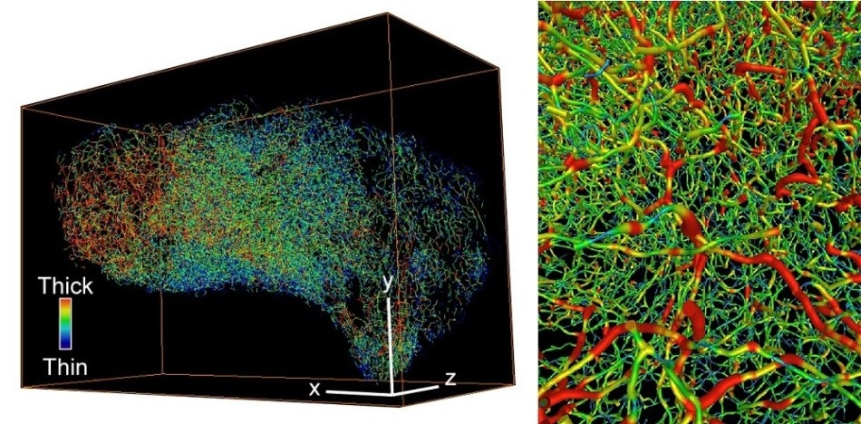
 3D images showing the complex vascular system in ovarian cancer (left) and bladder cancer (right). Thick vessels are colored red and thin vessels are colored blue. CREDIT: Images by Nobuyuki Tanaka.
Globally, on a daily basis, Pathologists investigate large samples of tumor tissues and the treatment for their patients depends on their diagnosis. However, in few instances, making the accurate diagnosis is highly difficult, meaning that wrong treatment is administered, which results in suffering of the patient and even death at times.
Prevalent techniques for pathological investigation for estimating the tumor stage involve adopting two-dimensional light microscopy. The stage of cancer indicates the growth and spread of the cancer, and is obligatory for administering the appropriate treatment. However, using two-dimensional techniques to investigate three-dimensional objects is not ideal and results in an information gap.
Hard to study 3D structures
To be sure, the tumors can be divided into sections for individual study, but three-dimensional structures such as the blood and lymph systems are very hard to study in this way.
Per Uhlén, Lead Investigator of the study and Professor, Department of Medical Biochemistry and Biophysics, Karolinska Institutet
The Scientists have adopted an innovative imaging method—a technique applied for basic research—to analyze human tumor tissue. In this method, the tissue is first made transparent. Then, it is reproduced in three dimensions in a so-called light-sheet microscope. The Scientists can use particular antibodies to label specific proteins to carry out a detailed analysis of the proteins.
“Light-sheet microscopy has been used in basic research for a while, but it is only in recent years that it’s been refined so much that it can be used practically in hospitals,” stated Professor Uhlén. “It was an unforgettable experience to look inside a tumor from a patient for the first time.”
Blood system structures give important information
The team collaborated with Clinicians from Karolinska University Hospital to analyze stored samples from patients who had bladder cancer. Then, apart from other structures, they recreated the three-dimensional blood system feeding the tumors in order to demonstrate that system can give detailed account of the aggressivity of the tumors. Using the innovative method, they could successfully and more precisely diagnose muscle-invasive tumors missed by two-dimensional techniques.
We’ve also studied other cancer types and judge that the method has considerable potential in the clinical diagnosis of all forms of solid tumors, especially cases that are difficult to diagnose. I hope that one day more pathological examinations will be conducted using 3D imaging.
Per Uhlén, Lead Investigator of the study and Professor, Department of Medical Biochemistry and Biophysics, Karolinska Institutet
Available at a KI core facility
The light-sheet microscope adopted to carry out this research is one among very few in Sweden and was obtained from the core facility Center for Live Imaging of Cells at Karolinska Institutet, or CLICK.
The Swedish Research Council, the Swedish Cancer Society, the Swedish Brain Foundation, the Knut and Alice Wallenberg Foundation, the Swedish Royal Academy of Science, the David and Astrid Hagelén Foundation, the Takeda Science Foundation, the Scandinavia-Japan Sasakawa Foundation and the Wenner-Gren Foundation funded this research.