Single-photon avalanche diodes (SPADs) operate as p-n junctions biased above their breakdown voltage. The field of laser scanning microscopy is undergoing rapid advancements, driven by the introduction of fast and compact detector arrays.
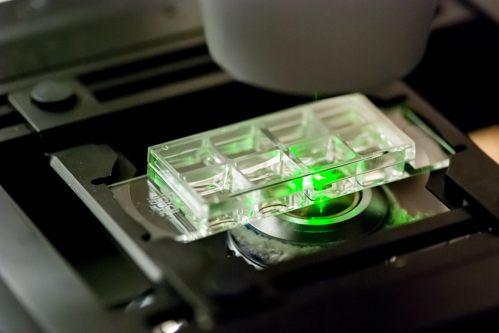
Image Credit: Micha Weber/Shutterstock.com
These detectors are gaining prominence in microscopy applications due to their exceptional reliability, robustness, ease of operation, and high detection efficiency. Asynchronous read-out SPAD array detectors are optimized to significantly enhance the capabilities of fluorescence laser-scanning microscopy (LSM). These specialized detectors provide distinctive single-photon spatiotemporal information, opening new possibilities for gentle and quantitative super-resolution imaging.
Introduction to SPAD Array Detectors
SPADs, utilized individually and in multi-pixel configurations, are primarily employed through single-photon counting (SPC), photon-timing facilitated by dedicated timing electronics, and time-gated SPC.
A recent article published in Frontiers in Physics highlights that when a photon is absorbed in the multiplication region of a SPAD, it can trigger a self-sustaining avalanche. This inherent positive feedback causes the rapid increase in current to a macroscopic level, enabling single-photon sensitivity and marking the arrival time of the detected photon with picoseconds time resolution. After each detection, a dead time is necessary to quench the avalanche and reset the SPAD, rendering the sensor temporarily blind.
Integrating SPADs and electronics on the same chip has enabled the natural progression of SPAD arrays, which involves incorporating the necessary auxiliary circuits for signal processing alongside the detectors. Various design strategies have been proposed to align with specific application requirements.
The initial stage involves an array of simple pixels, each comprising a SPAD and its frontend circuit. Subsequent stages involve integrating processing circuits into the pixel to execute specific functions, facilitating the parallel and independent operation of each SPAD pixel. Incorporating multiple SPADs and their frontend circuits within each pixel is also feasible to counteract the impact of the intrinsic SPAD dead time and enhance photon-number resolution.
SPAD-Enabled Handheld Fluorescence Lifetime Scanning Microscopy
Fluorescence imaging is a potent tool for material analysis, especially in biological applications, where numerous biomolecules display auto-fluorescence when illuminated.
In contrast to conventional fluorescence imaging, fluorescent lifetime imaging microscopy (FLIM) employs time-resolved detection systems to capture the characteristic fluorescent lifetime rather than solely focusing on the intensity or spectra of the emitted light.
In the realm of scanning microscopy, one of the most robust and elegant approaches for achieving the necessary time resolution is the utilization of SPADs.
In a recent article published in Optics Express, researchers demonstrated a handheld FLIM system employing a distally mounted SPAD array, occupying less than 2 mm2 with a resolution of 128 × 120, operating over a wired interface exceeding 1 meter in length. The research team used a specific camera called the Horiba FLIMera Time-Correlated Single Photon Counting (TCSPC) camera, which is commercially used for acquiring accurate fluorescent lifetime information.
The researchers presented an intense image of an ovine kidney captured using the handheld FLIM system. The image focused on the renal pelvis, where the ureter, veins, and arteries converge with the organ. In the fluorescence intensity image, there was no indication that the tissue in this region differed in composition from the rest of the organ.
The image exhibited notable contrast, with lifetimes ranging from approximately 1.2 ns to around 2 ns. Areas with very similar intensity levels displayed apparent differences in lifetime, aligning with previously published FLIM images of ovine kidney cross-sections.
The ability of the Endocam to provide FLIM images at a frequency exceeding 1 Hz while operating at a distance of approximately 1 m from its control board represents a significant achievement. This marks the first demonstration of a SPAD array operating in such a manner. These findings are useful as they confirm that the portable system can be used efficiently for bioengineering and other fields.
Challenges for SPAD Array Detectors
Most frontside-illuminated (FSI) SPAD detectors continue to exhibit relatively low fill factors—according to a recent research article published in Optics Express. Fill factor refers to the pixel area in the region of particle impact compared to the overall pixel area. Typically, in the case of SPADs, this factor is below 50 %. This limitation results in a restricted photon detection efficiency (PDE).
In scenarios with limited photons, particularly in bio-photonics, this can pose a challenge. Some studies have employed unique optical systems with SPAD acting as a special type of confocal pinhole, enabling the light to be directly focused on the photosensitive area. However, such setups are exceptions rather than the norm and often involve designs that are challenging to develop and maintain.
How Does Artificial Intelligence Improve SPAD Detector Imaging Systems?
Fluorescence lifetime imaging (FLI) is an imaging technique used for characterizing molecules based on the decay time from the excited state to the ground state.
Time-correlated single-photon counting (TCSPC) has gained popularity in FLI systems due to its advantages over other techniques in terms of time resolution, dynamic range, and robustness. In the past decade, SPADs have been successfully employed in TCSPC systems. However, current FLI systems have limitations in processing speed and accuracy.
Neural networks offer a novel avenue for rapid fluorescence lifetime extraction. In a groundbreaking study published in Scientific Reports, researchers have introduced the integration of a recurrent neural network (RNN) into SPAD-TCSPC systems for real-time FLI.
Unlike conventional deep learning methods that rely on histograms as input, which are available only post-data acquisition, RNNs eliminate histogramming and process raw timestamps in an event-driven manner. This approach facilitates incremental and continuous updating of lifetime estimations with every incoming photon, allowing for real-time or immediate post-acquisition readouts of fluorescence lifetime.
This innovative methodology obviates the need to store or transfer timestamp data or histograms, significantly alleviating the burden on hardware memory and data transfer requirements.
Breakthrough: Photon-Resolved Image Scanning Microscopy
Fluorescence confocal laser-scanning microscopy (LSM) is one of the most widely used tools in life science research. The growing popularity of LSM is anticipated to be further propelled by the emergence of single-photon SPAD array detectors specifically designed for LSM applications.
In contrast to conventional LSM single-element detectors, this class of sensors retains the spatial distribution of impinging fluorescence photons. Unlike a scientific camera, each array element operates as a fully independent SPAD, ensuring high temporal precision. Integrating modern data acquisition (DAQ) systems with these detectors enables efficient and fast photon-resolved measurements of emissions.
In a recent article published in Advanced Photonics, researchers successfully integrated a new Digital-Frequency-Domain (DFD)-based DAQ and control system into a single-photon laser-scanning microscopy (SP-LSM), incorporating a commercial SPAD array detector. This module implemented fluorescent lifetime imaging with super-resolution microscopy (FLISM).
The researchers used adaptive pixel reassignment (APR) to reconstruct a super-resolution image from the raw data.
FLISM demonstrated superior performance compared to conventional confocal laser scanning microscopy, exhibiting better resolution in the captured images. To further examine its viability, researchers measured living HeLa cells labeled with the CellBrite® NIR 680 cytoplasmic membrane dye.
FLISM provided insight into a wide distribution of fluorescence lifetime values, enabling the differentiation of plasma membranes (with longer fluorescence lifetime) from lipid-based structures or intracellular vesicles (with shorter fluorescence lifetime). The probe's fluorescence lifetime variation enabled effective monitoring of changes in the local environment.
The findings of this study indicate a strong connection between the future of laser scanning microscopy and SPAD array detectors. Integrating SPAD array detectors with a customized acquisition system simplifies access to and utilization of photon-resolved imaging spectroscopy microscopy.
More from AZoOptics: Surface Topography Imaging with AFM
References and Further Reading
SPIE. (2024). Building images photon-by-photon to increase the information content provided by microscopes. [Online] Phys.Org. Available at: https://phys.org/news/2024-02-images-photon-content-microscopes.html [Accessed 19 February 2024]
Cusini, I., et al. (2022). Historical Perspectives, State of Art and Research Trends of SPAD Arrays and Their Applications (Part II: SPAD Arrays). Frontiers in Physics. doi.org/10.3389/fphy.2022.906671
Matheson, A., et al. (2023). Handheld wide-field fluorescence lifetime imaging system based on a distally mounted SPAD array. Optics Express. doi.org/10.1364/OE.482273
Bruschini, C., et al. (2023). Challenges and Prospects for Multi-chip Microlens Imprints on front-side illuminated SPAD Imagers. Optics Express. doi.org/10.1364/OE.488177
Lin, Y., et al. (2024). Coupling a recurrent neural network to SPAD TCSPC systems for real-time fluorescence lifetime imaging. Sci Rep. doi.org/10.1038/s41598-024-52966-9
Tortarolo, G. et al. (2024). Compact and effective photon-resolved image scanning microscope. Advanced Photonics. doi.org/10.1117/1.AP.6.1.016003
Disclaimer: The views expressed here are those of the author expressed in their private capacity and do not necessarily represent the views of AZoM.com Limited T/A AZoNetwork the owner and operator of this website. This disclaimer forms part of the Terms and conditions of use of this website.