Researchers recently used cutting inspection techniques combining microscopy with spectroscopy to shed new light on millennia-old Egyptian papyrus, uncovering new information about this ancient historical period.
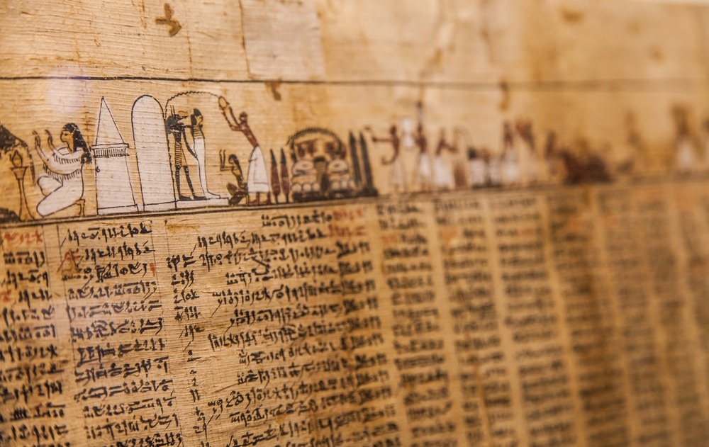
Image Credit: Paolo Gallo/Shutterstock.com
Illuminating Ancient Egyptian Illustration Practices
Illustrated papyruses have survived for millennia since they were created in ancient Egypt. These records depict stories and practices from the far past in vibrant colors and creative designs. France’s Musée Champollion recently allowed researchers from the country’s Université Grenoble Alpes, CNRS, and the European Synchrotron Radiation Facility in Grenoble to analyze its collection of ancient Egyptian papyrus. The Champollion’s papyruses show scenes from Egypt’s Book of the Dead, which contained instructions for preparing for the afterlife.
To examine these artifacts, the researchers employed several non-destructive inspection methods. They combined optical microscopy, Raman spectroscopy, synchrotron X-ray powder diffraction, and X-ray fluorescence.
They found that the usual illustration process from the ancient Egyptian New Kingdom period was used to make the papyruses. This confirmed previous conjecture that papyruses were illustrated with the same process that ancient Egyptians used to make mural paintings.
This process involves three steps: a preparatory red hematite drawing, coloring in with various pigments known to be used by ancient Egyptians, and a final black contour line drawing with carbon-based ink.
Analysis revealed that the final contour was sometimes significantly different from the preliminary sketch, which experts concluded was a sign of individual creativity used to make the papyrus. The researchers said that these results have increased knowledge about ancient Egypt’s illustration practices, proving that individual artistic intent was a factor in the final design of Egyptian records and decorations.
What Inspection Methods Were Used to Examine Ancient Egyptian Papyrus?
Two papyrus fragments were selected from the Champollion collection for inspection.
First, optical microscopy was used to investigate the two fragments. This was an open, exploratory investigation that provided data for the next step.
Using the optical microscopy data, scientists selected four regions from the two fragments for X-ray fluorescence spectrometry (XRF). In XRF, samples are bombarded with X-rays, and the resulting scatter is examined using spectrometry. It is used widely for elemental and chemical analysis, and as a non-destructive technique is widely applied in archaeology and forensic science.
XRF analysis revealed that iron, lead, copper, arsenic, and mercury were the main chemical elements relevant in the papyrus.
X-ray diffraction (XRD) was then used along four lines that crossed the four regions previously inspected with XRF. XRD is a powder diffraction technique that applies X-rays to powder or microcrystalline samples. It provides data on materials’ structural makeup by recording changes to the known wavelength of X-rays caused by interaction with different kinds of matter. The pattern of X-rays recorded on an XRD device’s sensor is then matched with patterns on a database to identify the presence of different materials. XRD is advantageous due to its reduced need for sample preparation and ability to analyze unknown materials quickly.
Using XRD and Rietveld and Pawley refinements, scientists identified hematite, cuprorivaite, atacamite, orpiment, realgar, and challacolloite in the pigments used to color the papyrus.
In a final step, the researchers used Raman spectroscopy to investigate the black pigment used to draw the outer, dark contour lines.
Raman spectroscopy is a technique for determining molecules' vibrational modes. It is commonly used to provide a fingerprint of molecules’ chemical structure, by which the molecule can be identified. It is also a non-destructive method for chemical identification and analysis, and as such is commonly used to investigate artifacts like paintings and manuscripts.
They found characteristic features of amorphous carbon throughout both fragments. This provided further evidence for previous findings by the researchers that black lines in Egyptian papyruses were made with flame carbon.
What Did the Scientists Discover About Ancient Egypt?
With the data from these analyses, scientists and historians were able to enhance our knowledge of ancient Egypt. Their important discovery was that papyrus was created using the same three-step process employed to create murals, and this enabled individual artists to express creativity while producing these artifacts.
Researchers highlighted what they interpreted as free and careful individual choices made by artists. Working within a set standard practice, they would have chosen how to color illustrations depending on pigments and shades available to them, adapting to individual scenes. It was also noted that artists would use adjuvants like driers to make particular colors adhere better.
This creativity also extended to the final step in papyrus production. Here, artists would draw freely, setting their individual creative stamp on the final look of the artifact.
Overall, the combination of optical microscopy, Raman spectroscopy, XRD, and XRF spectrometry led to an increase in our knowledge of ancient Egypt. We know more about how ancient craftspeople mass-produced and illustrated the “Book of the Dead,” a process now characterized by a subtle balance between creativity and standardization.
More from AZoM: Microscopy and Magnetic Characterization Analyzes London Underground Air Quality
References and Further Reading
Autran, P-O., et al (2023). Illustrating papyrus in Ancient Egypt. Scientific Reports. doi.org/10.1038/s41598-023-27761-7.
De Viguerie, L., V.A. Sole, and P. Walter (2009). Multilayers quantitative X-ray fluorescence analysis applied to easel paintings. Analytical and Bioanalytical Chemistry. doi.org/10.1007/s00216-009-2997-0.
Jung, D.S., et al (2018). High-energy X-ray applications: current status and new opportunities. Powder Diffraction. doi.org/10.1017/S0885715617001191
Disclaimer: The views expressed here are those of the author expressed in their private capacity and do not necessarily represent the views of AZoM.com Limited T/A AZoNetwork the owner and operator of this website. This disclaimer forms part of the Terms and conditions of use of this website.