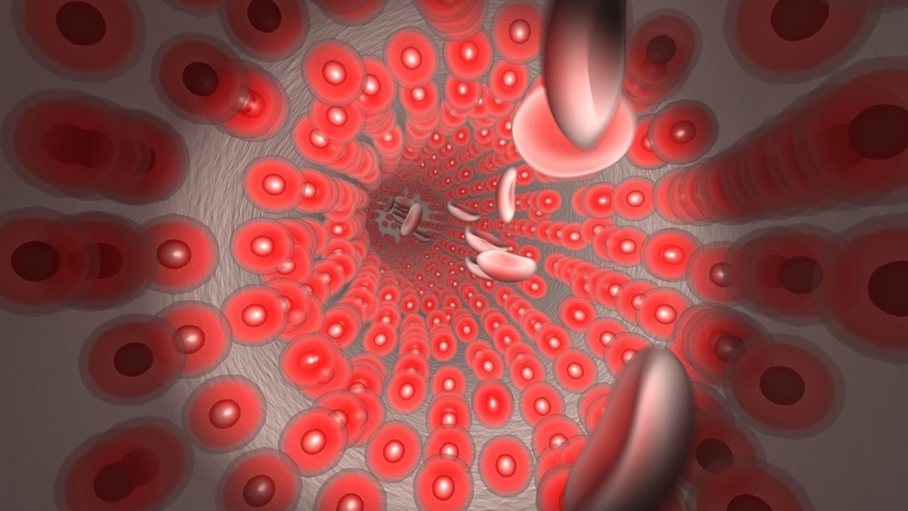
Fiber optic microendoscopes have become invaluable imaging tools that have enabled endoscopists to see and describe previously inconceivable things. This article will look into their history, applications, recent research and developments, challenges, and future industry prospects.

Image Credit: Daniel Reiner/Shutterstock.com
Fiber Optic Microendoscope: An Overview
The fiber optic microendoscope is based on fiber-optical confocal scanning microscopy, in which optical fibers are employed to transmit and collect signals. In addition, it uses an imaging lens at the probe end to generate high-resolution imaging.
Due to their excellent flexibility and small diameter, fiber optic microendoscopes allow valuable biomedical treatments like in vivo neuroimaging, microsurgery, and minimally invasive diagnostics.
Fiber-optical microendoscopy overcomes the physical constraints of conventional microscopy and enables less invasive and localized early cancer detection and microsurgery.
History of Fiber Optic Microendoscopes
Philipp Bozzini, a German physician, invented the Lichtleiter (light conductor) in 1806. This device employed candles, mirrors, and specula to investigate the esophagus, nose, and bladder. The technology continued to develop over the next decades, with advancements such as using lenses and little electric lamps for increased magnification at the instruments' tips.
Further advances came in the 20th century with the development of semiflexible devices and endoscopic tubes to transmit cold light to body cavities. Finally, in 1957, Larry Curtiss and Basil Hirschowitz developed the first fiber-optic endoscope, heralding the modern era of fiber-optical microendoscopy.
Applications
In Situ Cellular Imaging
Fiber optic micro-endoscope is a robust, versatile, and cost-effective approach for cellular imaging detail in situ, making it ideal for clinical and biomedical research. Micron-scale cells are imaged and optically stimulated in these fields at depths inaccessible to traditional microscopes.
Microendoscopy for Microsurgery
A fiber optical microendoscope was introduced for 3D in vivo imaging to regulate a femtosecond pulsed beam for microsurgery. The high-resolution 3D imaging provides detailed information, allowing the biopsy to be performed precisely to prevent complications.
Imaging Living Neurons
A multimode optical fiber microendoscope can produce a small fluorescent spot at one end, which can be moved to various locations on the tissue without shifting the fiber. Scanning the small spot over the sample excites fluorescent chemicals marking neuron activity. When the fluorescence from individual spots is sent back via the fiber, an image of neuronal activity is generated.
Challenges
Some fiber optic microendoscopes generate images by placing the probe directly against tissues, restricting imaging to the mucosa and surface epithelium. In addition, the mucosa cannot be sampled entirely by a fiber optic microendoscope; hence it is difficult to obtain accurate results, especially in areas of the body with varying tissue composition.

Image Credit: Roman Zaiets/Shutterstock.com
Recent Research and Development
Human Brain Imaging through a Multimode Fiber Optic Microendoscope
Vrije Universiteit Amsterdam researchers developed an optical fiber microendoscope as thin as human hair for minimally invasive deep brain tissue imaging to observe the impact of brain disorders such as Alzheimer's disease. The ultrathin multimode fiber optic microendoscope fits easily into an acupuncture needle, allowing for real-time imaging of deep tissues.
The research lays the foundation for non-invasive in vivo brain imaging and long-term neuronal activity monitoring.
Rice University's Fiber Optic Microendoscope Eliminates Unnecessary Biopsies
A portable, low-cost, battery-powered optical fiber microendoscope designed by Rice University bioengineers will remove the need for expensive biopsies for patients undergoing conventional endoscopic screening for esophageal cancer.
The microendoscope generates high-resolution images and real-time histological data, enabling endoscopists to view cell nuclei and individual cells in malignant tumors and rule out malignancy without a biopsy.
3D Optical Biopsies
Researchers from RMIT University in Australia produced microscopic 3D images of tissues using current optical fiber microendoscope technology.
The technique is based on the principles of light field imaging, in which multiple cameras view the same image from slightly different angles. In addition to medical applications, the microendoscopic imaging system can be employed in biological research for in vivo 3D fluorescence microscopy.
New Probe Design Improves Biomedical Imaging
Researchers from the Technical University of Denmark and the University of Western Australia demonstrated that side-viewing probes (microendoscopic imaging probes) combining spherical lenses and fiber-optic (GRIN) offer exceptional performance over a wide range of numerical apertures and enable a wider variety of imaging applications.
The novel design provides access to low-numerical aperture, long-working-distance applications with a smaller probe size and easier manufacturing. The increased working distance performance of the proposed design is especially promising for applications using a catheter- or needle-encased side-viewing probes.
Future of the Fiber Optic Microendoscopes
The future of fiber optic microendoscopy will be determined by the fast development of miniature lenses, optical fibers, and light sources that will enable 3D bioimaging in multiple spectral regimes.
The rapid development of this technology will result in a trend toward all-in-one imaging for microscopic and functional imaging, which will aid endoscopists in improving diagnostic accuracy, particularly the detection rate of malignant tumors.
Further advancements in automated analysis, usability, and functionality are expected to assist the technology's wide adoption and utilization. Therefore, numerous prominent research and business organizations with diverse approaches to fiber optic microendoscopy are devoting considerable resources to making the technology publicly accessible.
More from AZoOptics: The Future of Microscopic Tissue Imaging Using Ultra-Thin Endoscopes
References and Further Reading
Boas, G. (2013). The Recent History of Endoscope Design: Way More than Candlelight and Specula. [Online]. BioPhotonics. Available at: https://www.photonics.com/Articles/The_Recent_History_of_Endoscope_Design_Way_More/a55310 (Accessed on December 21 2022).
Gu, M., Bao, H., & Kang, H. (2014). Fibre‐optical microendoscopy. Journal of microscopy, 254(1), 13-18. https://doi.org/10.1111/jmi.12119
Karnowski, K., Untracht, G., Hackmann, M., Cetinkaya, O., & Sampson, D. (2022). Superior Imaging Performance of All-Fiber, Two-Focusing-Element Microendoscopes. IEEE Photonics Journal, 14(5), 1-10. https://doi.org/10.1109/JPHOT.2022.3203219
Lochocki, B., Verweg, M. V., Hoozemans, J. J., de Boer, J. F., & Amitonova, L. V. (2022). Epi-fluorescence imaging of the human brain through a multimode fiber. APL Photonics, 7(7), 071301. https://doi.org/10.1063/5.0080672
Orth, A., Ploschner, M., Wilson, E. R., Maksymov, I. S., & Gibson, B. C. (2019). Optical fiber bundles: Ultra-slim light field imaging probes. Science advances, 5(4), eaav1555. https://doi.org/10.1126/sciadv.aav1555
Protano, M. A., Xu, H., Wang, G., Polydorides, A. D., Dawsey, S. M., Cui, J., ... & Anandasabapathy, S. (2015). Low-cost high-resolution microendoscopy for the detection of esophageal squamous cell neoplasia: an international trial. Gastroenterology, 149(2), 321-329. https://doi.org/10.1053/j.gastro.2015.04.055
The Optical Society. (2018). Ultrathin endoscope captures neurons firing deep in the brain: New fiber-based endoscope, tested in mice, poised to bring new insights into brain function. [Online]. ScienceDaily. Available at: www.sciencedaily.com/releases/2018/03/180326110031.htm (Accessed on December 21 2022).
Wang, B., Zhang, Q., & Gu, M. (2020). Aspherical microlenses enabled by two-photon direct laser writing for fiber-optical microendoscopy. Optical Materials Express, 10(12), 3174-3184. https://doi.org/10.1364/OME.402904
Wang, B., Zhang, Q., Chen, X., Luan, H., & Gu, M. (2020). Perspective of fibre‐optical microendoscopy with microlenses. Journal of Microscopy. https://doi.org/10.1111/jmi.12977
Disclaimer: The views expressed here are those of the author expressed in their private capacity and do not necessarily represent the views of AZoM.com Limited T/A AZoNetwork the owner and operator of this website. This disclaimer forms part of the Terms and conditions of use of this website.