Microscopy is the workhorse of medical diagnostics. Histopathology, one of the most important medical fields, involves using microscopy to examine the often minute changes in tissue samples due to disease. Histopathology work is essential not just as a diagnostic tool but also for understanding some of the origins of disease for research into potential treatments.1
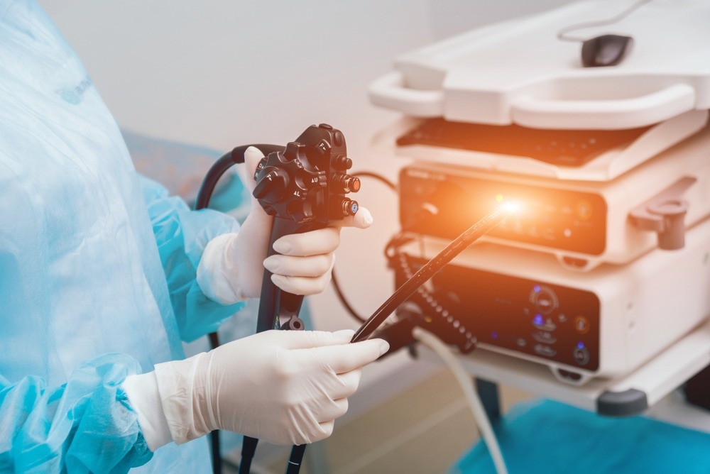
Image Credit: Roman Zaiets/Shutterstock.com
A wide range of microscopy methods is used for microscopic imaging of tissues, including Raman, optical light, and infrared microscopy.2-4
Some microscopy methods, such as Raman and infrared microscopy, help recover information on the chemical species present, adding an extra dimension for diagnosis. Others, such as optical microscopy, can be relatively straightforward to perform, which is essential in a busy hospital laboratory environment with a high rate of sample turnover.
Considerations when choosing an imaging method include which markers are considered ‘disease specific’, for example, whether the disease-induced changes at the cellular level are unique enough for a confident identification or whether additional chemical information on biomarker species is required.
Other considerations are the number of tissue samples available and their quality. Samples can degrade under poor storage conditions, making them harder to use for a confident diagnosis.5
Given that biopsies to obtain tissue samples are sometimes painful and have their own clinical risks, there have been recent developments to move microscopy methods from the laboratory setting to ‘in vivo’ measurements, where measurements are performed directly on the patient.
There are many potential advantages to using in vivo methods, including the ability for ‘real-time’ updates on patient status, such as checking that all cancerous tissues have been removed during surgery.6
What is Endoscopy?
One of the reasons for performing biopsies for histopathology is that surgical intervention is required to reach the site of the tumor or disease. One major advance in improving surgical outcomes has been the development of laparoscopic, sometimes known as keyhole, techniques. 7
When performing laparoscopic surgery, the surgical team needs to visualize the inside of the patient’s body while operating. To do this, devices known as endoscopes are used.
A standard endoscope is a thin tube-like device with intense light and a camera at the end. It is inserted into the site of interest, where it illuminates the tissue and transmits images through a fiber optic cable of the local region to a camera that the surgeon can monitor during the operation.
Endoscopes only require small incisions and are used as a surgical aid and a diagnostic tool in their own right.8 Most endoscopes use visible light sources and cameras, but it is also possible to use other sources and microscopy techniques.
Recent work has demonstrated the possibility of using a flexible, ultra-thin holographic endoscope for the microscopic imaging of tissues using an approach known as lensless Fourier holographic imaging.9
Holography Imaging
Holography experiments involve the reconstruction of a wavefront from a detected signal. With the advent of digital holography methods, this is often carried out algorithmically by a computer, where the intensity of the signal and phase information is reconstructed.
One advantage of holography methods for endoscopy is that they do not require lenses to achieve the exact spatial resolution as a standard transmission or fluorescence-based optical methods.
Lenses are generally bulky and endoscopes, where the fiber optical bundle needs to be as small as possible to minimize patient discomfort, are not always practical. Using fiber bundles to improve the light collection efficiency is undesirable as this adds to the probing instrument's bulk.
With the holographic endoscope, it was possible to achieve a lateral and axial spatial resolution of 0.85 μm and 14 μm with a fiber bundle less than < 350 μm in diameter.9 The team also stated that further reducing the bundle size would be very straightforward.
Future Possibilities of Endoscopy Techniques
Compared to lab-based optical microscopy methods that often require staining or pre-treatment of samples, the endoscope design is advantageous as no preparation of the tissue for imaging is needed.
The team achieved very high contrast in the images, making it easier to identify objects.
Further improvements include making the algorithms that perform the image processing more efficient to help reduce motion artifacts in the images.
The endoscope used was also flexible, making it easier to insert. As the new endoscope is comparable in size to an acupuncture needle, it could also be inserted into regions such as the tiny airways in the lung and even potentially into the brain.
With adaptations to the device, the team also suggests it may be possible to use this for light-based medical treatments, where laser pulses are sent down the fiber to irradiate tumors in vivo or light is used to photoactivate specific pharmaceutical compounds.
References and Further Reading
- Musumeci, G. (2014). Past, present and future: overview on histology and histopathology. Journal of Histology and Histopathology, 1(1), 5. https://doi.org/10.7243/2055-091x-1-5
- Mittal, S., Yeh, K., Suzanne Leslie, L., Kenkel, S., Kajdacsy-Balla, A., & Bhargava, R. (2018). Simultaneous cancer and tumor microenvironment subtyping using confocal infrared microscopy for all-digital molecular histopathology. Proceedings of the National Academy of Sciences of the United States of America, 115(25), E5651–E5660. https://doi.org/10.1073/pnas.1719551115
- Mittal, S., Wrobel, T. P., Walsh, M., Kajdacsy-Balla, A., & Bhargava, R. (2021). Breast cancer histopathology using infrared spectroscopic imaging: The impact of instrumental configurations. Clinical Spectroscopy, 3(July 2020), 100006. https://doi.org/10.1016/j.clispe.2021.100006
- Polli, D., Kumar, V., Valensise, C. M., Marangoni, M., & Cerullo, G. (2018). Broadband Coherent Raman Scattering Microscopy. Laser and Photonics Reviews. 1800020. https://doi.org/10.1002/lpor.201800020
- Ladekarl, M. (1994). The influence of tissue processing on quantitative histopathology in breast cancer. Journal of Microscopy, 174, 93–100. https://doi.org/10.1111/j.1365-2818.1994.tb03453.x
- Wells, W. A., Thrall, M., Sorokina, A., Fine, J., Krishnamurthy, S., Haroon, A., Rao, B., Shevchuk, M. M., Wolfsen, H. C., Tearney, G. J., & Hairi, L. P. (2018). In Vivo and Ex Vivo Microscopy. Arch Pathol Lab Med, 143, 288–298. https://doi.org/10.5858/arpa.2018-0298-RA
- Okholm, C., Goetze, J. P., Svendsen, L. B., Patrick, M., Okholm, C., Goetze, J. P., Svendsen, L. B., Patrick, M., Okholm, C., Goetze, J. P., Svendsen, L. B. O., & Achiam, M. P. (2014). Inflammatory response in laparoscopic vs . open surgery for gastric cancer cancer. Scandinavian Journal of Gastroenterology, 49, 1027–1034. https://doi.org/10.3109/00365521.2014.917698
- Kuipers, E. J., & Haringsma, J. (2005). Diagnostic and Therapeutic Endoscopy. Journal of Surgical Oncology, 92, 203–209. https://doi.org/10.1002/jso.20361
- Choi, W., Kang, M., Hong, J. H., Katz, O., Lee, B., Kim, G. H., Choi, Y., & Choi, W. (2022). Flexible-type ultrathin holographic endo- scope for microscopic imaging of unstained biological tissues. Nature Communications, 13, 4469. https://doi.org/10.1038/s41467-022-32114-5
Disclaimer: The views expressed here are those of the author expressed in their private capacity and do not necessarily represent the views of AZoM.com Limited T/A AZoNetwork the owner and operator of this website. This disclaimer forms part of the Terms and conditions of use of this website.