Image Credit: Micha Weber/Shutterstock.com
Fluorescence microscopy has been in development for well over a century. The term fluorescence was originally coined in 1852 by British scientist Sir George S Stokes. Fluorescence microscopy technology, which provides enhanced contrast images at the resolution of the single-molecule, was influenced by the work of pharmacology professor Philipp Ellinger whose work in the 1920s helped establish the intravital fluorescence microscope. Over the years, research from keen scientists worldwide has contributed to the development of fluorescence technology and has helped to establish it as a reliable and dependable tool across various industries.
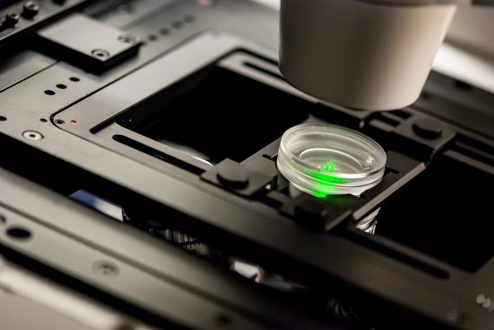
How Does Fluorescence Microscopy Work?
The fluorescence microscope produces ultraviolet light from either a mercury or xenon lamp. The ultraviolet light emitted from the microscope is reflected by a dichroic mirror, which directs it to the specimen.
Fluorescence within molecules of the sample is excited by the photons of fluorescent light, which is then collected by an objective lens and passed back through the dichroic mirror as well as a filter that prevents other wavelengths of light from passing through. The light then moves through to the eyepiece of the microscope to produce an image.
Fluorescent molecules can exist naturally within a sample, or they can be introduced by methods that stain cells with a dye. Different dyes are used to visualize living and dead cells; calcein/AM causes living cells to fluoresce green and propidium iodide causes dead cells to fluoresce red.
Fluorescence microscopy is useful for visualizing biological structures and observing biochemical and physiological events. DNA, calcium, magnesium, sodium, and enzymes have all been successfully studied with fluorescence microscopy. techniques that attach antibodies to sub-cellular structures have been developed to visualize intracellular activity as it happens.
What Industries is Fluorescence Microscopy Used In?
Because fluorescence microscopy can visualize biological structures both living and dead, the technique has become well established in the field of scientific research. It has also made a footprint in pharmaceuticals, to visualize how certain therapeutics may be impacting the body. It has also become important to medical diagnostics and disease monitoring.
The imaging technique is frequently used in industry as a method of quality control. The materials industry uses fluorescence microscopy to evaluate the internal structure and integrity of materials produced. The food industry uses the technique for quality control of food products.
What Key Research Has Been Conducted Using Fluorescence Microscopy?
Fluorescence microscopy has facilitated many key discoveries and advancements of knowledge in the pathophysiology of many diseases. The technique gives scientists a unique look into the molecular basis of disease, helping to develop enhanced methods of diagnosis and therapy as well as establish preventative strategies.
Oncology Advancements
Fluorescence microscopy has helped to make key advancements in oncology. Studies using this technique have uncovered cell adhesion dynamics and tumor cell migration. Recent advances within the field of fluorescence microscopy have further benefited oncology, with the development of recombinant fluorescent proteins (FPs), helping to enhance imaging of protein distribution, dynamics, and interaction within living cells at unprecedented spatial and temporal resolutions. As a result, the molecular mechanisms underpinning cell migration in cancer are now further understood.
Infectious Diseases
Infectious diseases have also greatly benefited from fluorescence microscopy. Again, the technique has facilitated the elucidation of the molecular mechanisms and host-pathogen interactions responsible for the establishment and progression of infectious diseases. Similarly, the imaging technique has advanced our knowledge of the biological mechanisms underlying rare genetic diseases.
Alzheimer’s Disease
Fluorescence microscopy has helped to develop our understanding of Alzheimer’s disease. The Aβ plaques and tau proteins that build up in the brain can be readily stained with thioflavins for fluorescent microscopic imaging of brain tissues. This has facilitated many advancements in our knowledge of the rare disease, for which there is currently no cure.
Other rare genetic diseases have benefited from fluorescence microscopy. Recently, research using the imaging technique has revealed the ultrastructure of the accumulations of the Huntingtin’s protein within neurons, which eventually results in neuronal atrophy.
Limitations of Fluorescence Microscopy
Like other forms of microscopy, fluorescence microscopy faces three general limitations: resolution, noise, and optical aberrations. However, as technology advances, these limitations are gradually being overcome.
In addition, fluorescence microscopy faces two specific limitations. First, it is dependent on the deposition of energy into the sample. Second, its use of exogenous fluorescent probes is not ideal as they can impact the cellular structure and function.
What Does the Future Hold for Fluorescence Microscopy?
The continued development of fluorescence microscopy will likely happen alongside developments in life sciences. The needs of scientific research will likely guide future advancements in capabilities of the imaging technique, which will fuel further advancements in scientific research, namely, our understanding of the disease at the molecular level.
References and Further Reading
Jun, Y., Cho, S., Jung, J., Huh, Y., Kim, Y., Kim, D. and Ahn, K., 2019. Frontiers in Probing Alzheimer’s Disease Biomarkers with Fluorescent Small Molecules. ACS Central Science, 5(2), pp.209-217. https://pubs.acs.org/doi/10.1021/acscentsci.8b00951
Le Dévédec, S., Yan, K., de Bont, H., Ghotra, V., Truong, H., Danen, E., Verbeek, F. and van de Water, B., 2010. Systems microscopy approaches to understand cancer cell migration and metastasis. Cellular and Molecular Life Sciences, 67(19), pp.3219-3240. https://www.ncbi.nlm.nih.gov/pmc/articles/PMC2933849/
Masters, B., 2010. The Development of Fluorescence Microscopy. eLS,. http://www.fen.bilkent.edu.tr/~physics/news/masters/ELS_Hist_Fl_Micro.pdf
Renz, M., 2013. Fluorescence microscopy-A historical and technical perspective. Cytometry Part A, 83(9), pp.767-779. https://onlinelibrary.wiley.com/doi/full/10.1002/cyto.a.22295
Sanderson, M., Smith, I., Parker, I. and Bootman, M., 2014. Fluorescence Microscopy. Cold Spring Harbor Protocols, 2014(10), p.pdb.top071795. https://www.ncbi.nlm.nih.gov/pmc/articles/PMC4711767/
Disclaimer: The views expressed here are those of the author expressed in their private capacity and do not necessarily represent the views of AZoM.com Limited T/A AZoNetwork the owner and operator of this website. This disclaimer forms part of the Terms and conditions of use of this website.