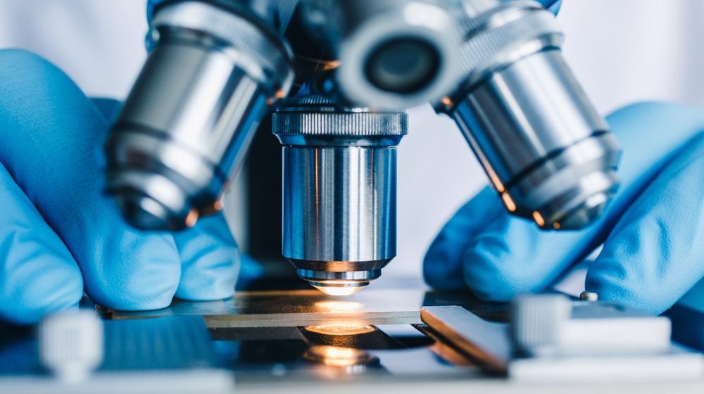
The field of microscopy has undergone several major advances in recent years.1 From the proliferation of super-resolution techniques that acquire sub-diffraction limit spatial resolution to improvements in wide-field imaging capabilities, it is now possible to use microscopy on an ever-growing range of samples and length scales.

Image Credit: Konstantin Kolosov/Shutterstock.com
The ability to visualize objects as small as viruses or particles has meant microscopy techniques have become an embedded part of quality control, medical diagnosis, and biological research. However, despite this wealth of advancement, there are still many challenges and limitations to be overcome with improvements in the technology and procedures for microscopy experiments.
Light Doses
One significant challenge in microscopy is photobleaching. Many microscopy methods, including most super-resolution approaches, make use of fluorescent labels for looking at samples. Fluorescent labels are molecules that interact with parts of a cell or substrate and have very high fluorescence quantum yields. Ideally, the specificity of such a label is high enough that the tag can be used to identify particular cell structures as these will be the only regions in which is bound and activated.
Many microscopy schemes make use of some sort of excitation process that targets these bound fluorophores. The excitation scheme in a microscopy experiment usually makes use of a laser source and, particularly for multiphoton microscopy methods, this means a significant amount of energy can be transferred to the sample on excitation.2
The wavelength of the laser is chosen so that it is resonant with an absorption band of the fluorescent tag. Once the label has been photoexcited, it will begin to emit light for a given period, defined by the emission lifetime of the tag. This light can then be collected to visualize the sample and any structures of interest.
The reason fluorescent labels are so commonly used in fluorescence, confocal, and super-resolution microscopy, is that their high fluorescence quantum yield makes them significantly easier to detect. Super-resolution techniques involve single-molecule imaging and so there will only be a small number of fluorophores that can be detected. No image can be recorded if each of these does not emit sufficient light to exceed the detection limits of the cameras used.
If signal levels are low, then one way to improve the signal-to-noise in a microscopy image is to increase the acquisition time. Depending on the lifetimes of the fluorescence tags used, longer acquisitions may require re-excitation of the fluorescent species or longer total illumination times. Both scenarios increase the light dose received by the sample and increase the probability of photobleaching.3
Photobleaching
Photobleaching occurs when the fluorescent label can no longer emit light, making it functionally useless. Photobleaching is problematic for several reasons. Firstly, if all the fluorophores have gone dark, there is no way to visualize certain regions of the cell. Secondly, photobleaching effects can interfere with kinetics measurements that are used extensively in live-cell imaging.3 If emission levels decrease due to degradation of the fluorophores rather than a biological process, false rate constants can be acquired. Photobleaching can also lead to changes in the physiology of the cells being measured.4
It is, therefore, desirable to minimize photobleaching effects. When designing new fluorophores, stability with respect to photobleaching is often a desirable property. Microscopy experiments can also be adapted to reduce photobleaching through modification of experimental conditions. This includes a reduction of the excitation intensities used and overall exposure times or the use of filters to attenuate certain incident wavelengths.
Adaptive Microscopy
An alternative approach is to make use of adaptive microscopy methods.5 As microscopy techniques have been developed to scan larger sample areas, it is challenging to avoid photobleaching due to increased exposure time.
With adaptive microscopy platforms, such as the instrument recently developed in the Institut Fresnel at Aix Marseille University, France, scan times can be reduced by performing more efficient scan patterning over the sample. This can be achieved because many biological structures are part of regular, well-characterized architectures.
For example, many types of organs and embryos are made of cells organized as a surface that forms a curved shape. These types of structures tend to be particularly delicate with respect to photobleaching and the regions that are generally of most interest are parts of the structure that lie at the cell periphery.
In a traditional scanning fluorescence microscopy experiment, the entire organ would be systematically scanned but by combining the image output with a live feedback system consisting of image recognition and scan control, the team minimized the scan time over regions of less interest.
The use of this feedback system resulted in a reduction in the overall light exposure by up to a factor of 80 in comparison to full scans of the object volume. The team has also made use of a similar approach for imaging living organisms.6
Adaptive scanning has the potential to be integrated with existing microscopy set-ups and for more effective scanning of precious samples, offering both timesaving and artifact suppression from photobleaching minimization.
References and Further Reading
- Nimesh, N., Joon, D., & Nimesh, M. (2017). Advances in Microscopy-A Review. Voyager: Vol.III, 1, 0976–7436.
- Gavin, R. H. (2009). Cytoskeleton Methods and Protocols.
- Goldberg, Y., Ashby, M. C., & Henley, J. M. (2012). Measuring Membrane Protein Dynamics in Neurons Using Fluorescence Recovery after Photobleach. In Methods in Enzymology (Vol. 504, pp. 127–146). https://doi.org/10.1016/B978-0-12-391857-4.00006-9
- Carlton, P. M., Boulanger, J., Kervrann, C., Sibarita, J. B., Salamero, J., Gordon-Messer, S., Bressan, D., Haber, J. E., Haase, S., Shao, L., Winoto, L., Matsuda, A., Kner, P., Uzawa, S., Gustafsson, M., Kam, Z., Agard, D. A., & Sedat, J. W. (2010). Fast live simultaneous multiwavelength four-dimensional optical microscopy. Proceedings of the National Academy of Sciences of the United States of America, 107(37), 16016–16022. https://doi.org/10.1073/pnas.1004037107
- Abouakil, F., Meng, H., Burcklen, M., & Rigneault, H. (2021). An adaptive microscope for the imaging of biological surfaces. Light: Science & Applications. https://doi.org/10.1038/s41377-021-00649-9
- Royer, L. A., Lemon, W. C., Chhetri, R. K., Wan, Y., Coleman, M., Myers, E. W., & Keller, P. J. (2016). Adaptive Light-sheet microscopy for long-term, high resolution imaging in living organisms. Nature Biotechnology, 34(12), 1267. https://doi.org/10.1038/nbt.3708
Disclaimer: The views expressed here are those of the author expressed in their private capacity and do not necessarily represent the views of AZoM.com Limited T/A AZoNetwork the owner and operator of this website. This disclaimer forms part of the Terms and conditions of use of this website.