The most common tests for detecting SARS-CoV-2 infections have become a combination of antigen rapid lateral flow tests and reverse transcriptase-polymerase chain reaction (RT-PCR) of swabbed material from the patient. While RT-PCR tests are typically considered to be more accurate and reliable than antigenic tests, the laboratory time required to develop and analyze samples is significant, particularly when a rapid diagnosis is required.
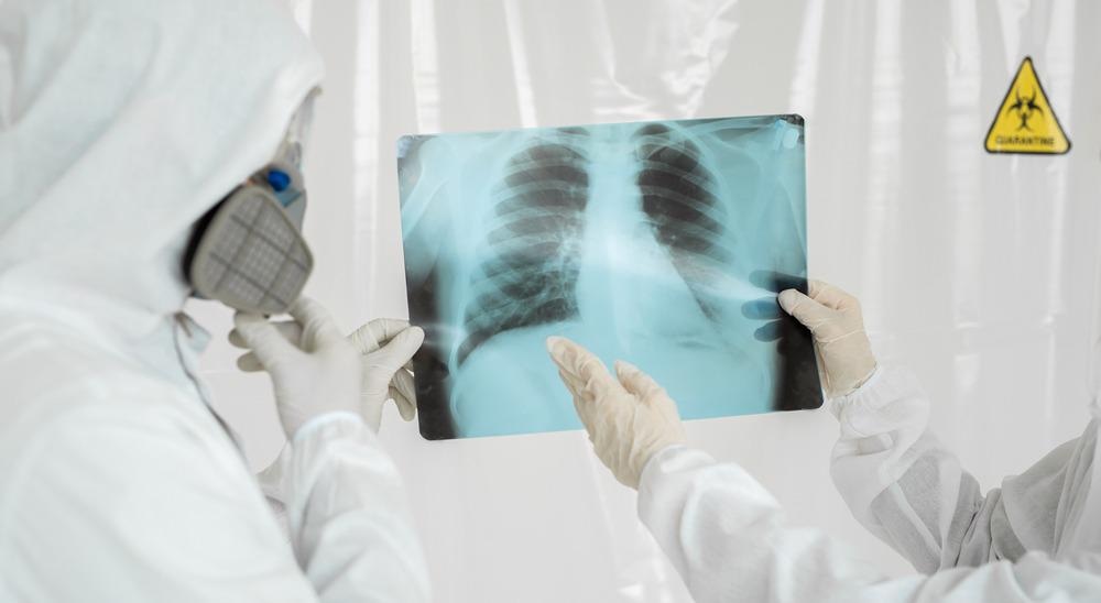
Image Credit: Studio Peace/Shutterstock.com
As COVID-19 is a respiratory disease, one of the key areas affected is the lungs. Mild cases can lead to inflammation and irritation, with more serious cases leading to extreme inflammation, damage to the alveoli, and filling of the lungs with fluid (pneumonia). Inflammation and lung damage of this type can affect the appearance of the tissue in the lungs, making regions appear to be fused together, and fluids can give chest images a milky, or ground glass, appearance.
Studies Investigating the Diagnosis of COVID-19 via Imaging
Due to the prevalence of respiratory symptoms as part of COVID-19, recent studies have been investigating using imaging techniques, such as chest computed tomography (CTs), X-rays, and ultrasound, as rapid, accurate diagnostics of COVID-19 disease.1 In response to this, the World Health Organization (WHO) has collated a rapid review of the effectiveness of medical imaging approaches to COVID-19 diagnosis. This is to help clinicians and researchers alike rapidly assess new and developing approaches for improved patient outcomes.
Although currently the WHO’s recommendations on the use of imaging for COVID-19 diagnosis are conditional as they are still based on low-certainty evidence, this recommendation may change as further studies are completed.2
One of the largest systematic reviews on COVID-19 diagnosis using imaging to date has been the Cochrane systematic review of 51 independent studies. This systematic review compared the accuracy and specificity of three main types of medical imaging for the thoracic region, including ultrasound, chest CTs, and 2D X-ray imaging.
Of the imaging technique trialed, ultrasound is the most easily accessible and can be performed in a local doctor’s office. CT scans however require access to highly specialized equipment and trained staff for operation and analysis, though provide 3D reconstructions of multiple 2D X-ray images.
From the results of the systematic review looking at different studies attempting to use standard 2D X-ray images to diagnose COVID-19, it was identified that a 2D chest X-ray was sufficient to correctly diagnose COVID-19 in 80.6% of infected patients.1 However, the study also reported a 28.5% false-positive rate.
In comparison to single 2D X-ray images, chest CTs offered a greater degree of diagnostic accuracy, correctly identifying positive COVID-19 cases with 87.9% accuracy, but incorrectly identifying COVID-19 in 20% of patients who did not have COVID-19.
While lung ultrasounds offered good results in correctly diagnosing 86.4% of cases where the patient had COVID-19, the false positive rate was significantly higher than the other two diagnostic methods, with 45% of people being incorrectly identified as suffering from COVID-19.
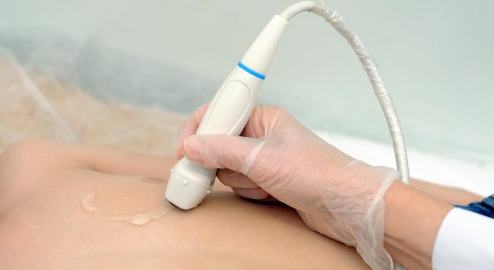
Ultrasounds have been used to detect COVID-19 in patients. Image Credit: LIAL/Shutterstock.com
Part of the challenge of comparing these different imaging studies is that, as COVID-19 is still a new disease, there is no universally agreed definition of positivity. This means that there is no agreed-upon set of features that could be identified in an image that would be strongly correlated with COVID-19 infection and would contribute to both false negative and positive errors. For example, a cloudy or milky image could be a sign of pneumonia as a result of another respiratory illness, not just COVID-19, so a good definition of positivity would include symptoms more unique to COVID-19.
However, while the WHO still advise an RT-PCR test as the most accurate and preferred method of diagnosis, when results are delayed or access to laboratory tests is not possible, for patients with mild to moderate suspected COVID-19, the WHO still considers that imaging techniques can be a useful tool to guide therapeutic management. The patients most likely to benefit from this are those with a high risk of disease progression or are already unresponsive to treatment.
The preliminary suggestions from the systematic review are that chest radiography may offer higher specificity, if reduced sensitivity, than a chest CT. The significant advantage being that recording 2D X-rays is significantly less resource-intensive and, as the radiation dose is smaller, disease progression can also be monitored using this imaging technique.
The Future of Using Imaging Methods to Detect COVID-19
Imaging techniques as diagnostic tools for COVID-19 may offer significant advantages over RT-PCR as such tests are often poor at diagnosing the earliest stages of the disease. Early intervention is also key to improving the prognosis. Therefore, refining diagnostic imaging techniques and further improving their accuracy and specificity is an essential part of improving COVID-19 outcomes.
References and Further Reading
- Islam N, Ebrahimzadeh S, Salameh J-P, Kazi S, Fabiano N, Treanor L, Absi M, Hallgrimson Z, Leeflang MMG, Hooſt L, van der Pol CB, Prager R, Hare SS, Dennie C, Spijker R, Deeks JJ, Dinnes J, Jenniskens K, Korevaar DA, Cohen JF, Van den Bruel A, Takwoingi Y, van de Wijgert J, Damen JAAG, Wang J, McInnes MDF, Cochrane COVID-19 Diagnostic Test Accuracy Group. (2021) Thoracic imaging tests for the diagnosis of COVID-19. Cochrane Database of Systematic Reviews, Issue 3. Art. No.: CD013639. DOI: 10.1002/14651858.CD013639.pub4.
- Akl, E. A., Blažić, I., Yaacoub, S., Frija, M. P. H. G., Chou, R., Jin, Z., … Jane, P. (2021) Use of Chest Imaging in the Diagnosis and Management of COVID-19: A WHO Rapid Advice Guide. Radiology, 298(2), E64-69. https://doi.org/10.1148/radiol.2020203173
Disclaimer: The views expressed here are those of the author expressed in their private capacity and do not necessarily represent the views of AZoM.com Limited T/A AZoNetwork the owner and operator of this website. This disclaimer forms part of the Terms and conditions of use of this website.