Surface Enhanced Raman Spectroscopy (SERS) is a powerful technique that is used to enhance sensitivity when using Raman spectroscopy. Raman scattering gives inherently weak signals, but the discovery of SERS in the 1970's1 by Martin Fleischmann and his team from the University of Southampton, has helped to overcome this issue.
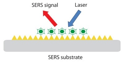
Illustration of how SERS works. (Image credit: Semrock)
What is it?
SERS was first discovered in 1974 when a Raman spectra of pyridine on roughened silver was observed, but the impact of this discovery was not further realized until 1977. Typical metals used are gold, silver and copper, but research into using the alkali metals and metal alloys as plasmonic substrates for SERS has also been explored. The roughened surfaces are prepared through chemical roughening, metallic coating, or deposition of colloidal metallic nanoparticles on to a surface, and a laser wavelength compatible with the chosen SERS metal is used for excitation2.
Laser excitation of the nanostructures creates a plasmonic light field. When a molecule lies close to the enhanced field at the surface or is absorbed, an enhancement in the Raman signal is observed. The enhanced signals enable the detection of low concentrations, so fluorescence and labeling are not required2.
How is it used?
SERS is used in a wide range of fields, including forensics, medical testing, analytical testing, trace material analysis, drug discovery, point of care testing, and biological and chemical threat detection. The potential uses for SERS in biosensing are vast and include detection of diseases such as diabetes, cancer, Alzheimer’s and Parkinson’s.
Immunofluorescence staining is used for clinical diagnosis of tissues, but the toxicity of various fluorescent dyes means that analysis cannot be carried out in vivo. Raman spectroscopy is a label-free diagnostic technique thta is chemically specific, so is a great alternative3. The fact that SERS is a fast, label-free technique has also proved to be a great advantage for point-of-care tests for therapeutic drug monitoring4.
SERS nanotags, gold spheres functionalized with reporter molecules encased in a silica shell, can be used as a way of labeling and authentication of different objects. The nanotags can be used not just to encode jewelery and banknotes for security, they can also be used for identification of fraud during transportation of goods2.
With Raman and SERS being made portable, the use of SERS in field testing for forensics and biological and chemical threat detection has greatly increased. The need for portable detection equipment is great, with previous methods of analysis such as using liquid chromatography and mass spectrometry requiring lengthy sample preparation, and SERS providing a great, sensitive and fast alternative2.
Type of SERS Surfaces
One of the most common methods of SERS enhancement is by using metallic nanoparticle solutions. The nanoparticle solutions normally consist of gold and silver colloid, but research into advancing SERS technology has led to a wide variety of shapes, sizes and coatings of the nanoparticles.
Gold and silver nanospheres are the most common nanoparticle types used in SERS, but other shapes such as nanostars, nanorods, nanocubes and nanowires can be produced through a polymer-mediated polyol process. Nanoparticles can also be capped or hollowed using various chemical methods. Both the shape and size of nanoparticles have been shown to affect SERS enhancement5. As well as direct detection with samples mixed with nanoparticles, these nanoparticles can be deposited or spin-coated onto various surfaces for a more accurate spread for detection, similar to using solid roughened surfaces.
Solid roughened solid surfaces can be made through nanolithography, but nanolithography is expensive compared to common solution-based nanoparticle methods, so it is a less common use for SERS substrates. There are a few commercially available solid SERS substrates, such as Klarite, which is a substrate comprised of pyramidal shaped pits etched into silicon. A gold/silver nanosponge by Ocean Optics is also available, as well as multiple gold and silver substrates from Silmeco. There is a lot of research being done by companies such as the DSTL and Universities such as The University of Strathclyde into making new SERS substrates that would be alternatives to those currently commercially available.
New Research
SERS remains an area of analytical chemistry that still has many areas left to be explored to get the best results possible. Researchers are developing both new ways of using nanoparticles for SERS enhancement, as well as looking into how different solid roughened surfaces could enhance SERS signals.
As well as improving current methods of detection, SERS has the possibility in the future to be combined with other techniques. Combining ultraviolet spectroscopy with SERS would allow for the detection of proteins and biomolecules. Ultrafast SERS could be possible if it is able to be combined with femtosecond stimulated Raman spectroscopy. There is also a chance that tip-enhanced Raman spectroscopy is possible in the future, but a lot of challenges need to be overcome first before that would be possible2.
With a lot of spectroscopy research groups now looking into SERS technology, there is a lot of hope for the future of SERS, but a lot more research left to be done.
References:
- M. Fleischmann, P. J. Hendra, A. J. McQuillan, Raman spectra of pyridine adsorbed at a silver electrode. Chemical Physics Letters. 1974 DOI: 10.1016/0009-2614(74)85388-1
- Bhavya Sharma, Renee R.Frontiera, Anne-Isabelle Henry, Emilie Ringe, Richard P.Van Duyne, SERS: Materials, applications, and the future, Materials Today. 2012 DOI: 10.1016/S1369-7021(12)70017-2
- Sebastian Wachsmann-Hogiu, Tyler Weeks, Thomas Huser, Chemical analysis in vivo and in vitro by Raman spectroscopy – from single cells to humans, Current Opinion in Biotechnology. 2009 DOI: 10.1016/j.copbio.2009.02.006
- Aleksandra Jaworska, Stefano Fornasaro, Valter Sergo, Alois Bonifacio, Potential of Surface Enhanced Raman Spectroscopy (SERS) in Therapeutic Drug Monitoring (TDM). A Critical Review, Biosensors. 2016 DOI: 10.3390/bios6030047
- Pamela A. Mosier-Boss, Review of SERS Substrates for Chemical Sensing, Nanomaterials. 2017 DOI: 10.3390/nano7060142
Disclaimer: The views expressed here are those of the author expressed in their private capacity and do not necessarily represent the views of AZoM.com Limited T/A AZoNetwork the owner and operator of this website. This disclaimer forms part of the Terms and conditions of use of this website.