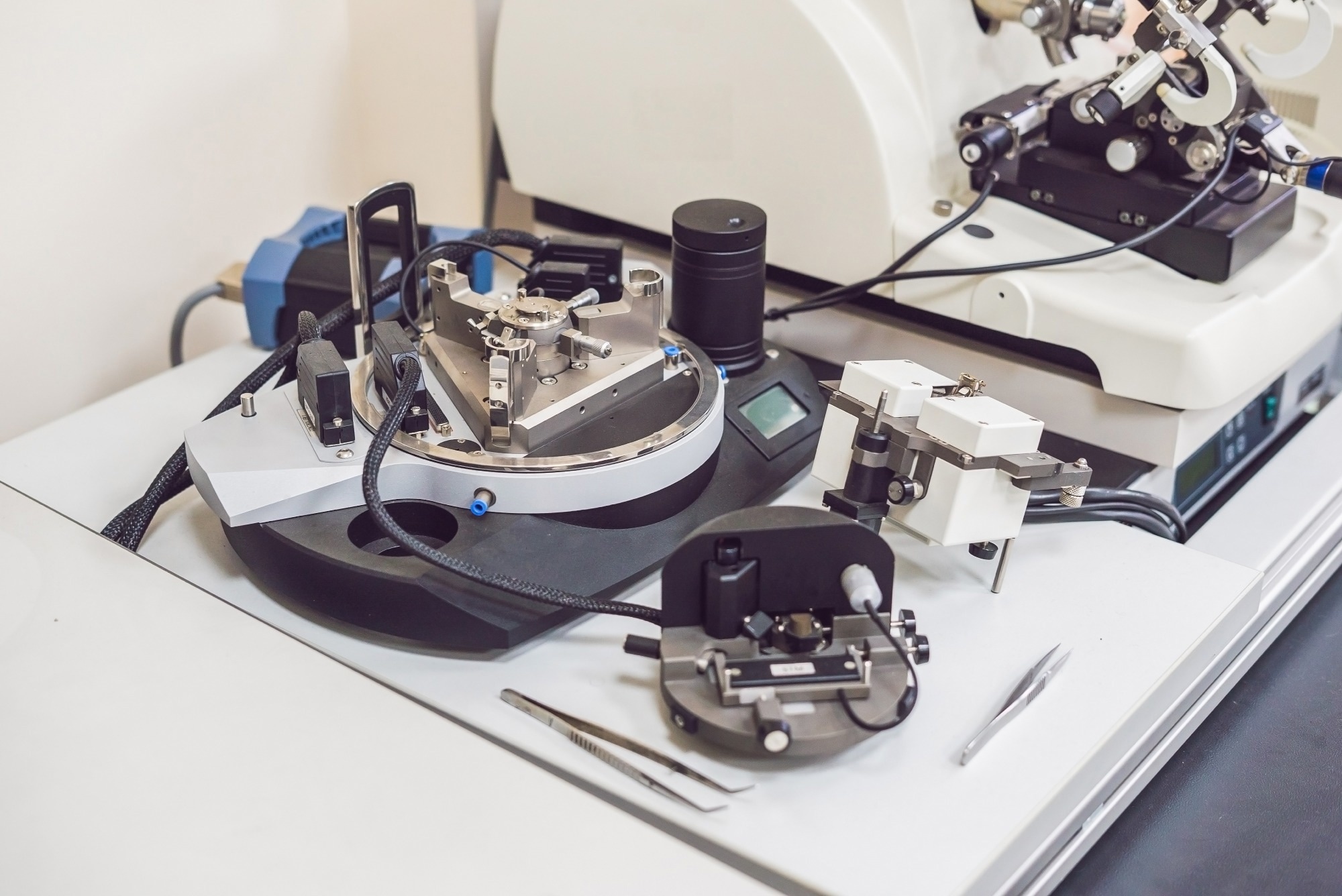
A recent study published in Advanced Photonics Research explores how refining atomic force microscopy (AFM) techniques can improve high-resolution imaging of human corneal epithelial cells (HCET).
The researchers focused on boosting optical detection sensitivity and image quality in liquid environments to better visualize the native structure of soft biological samples.

Image Credit: Elizaveta Galitckaia/Shutterstock.com
The Role of AFM in Nanoscale Imaging
Since its introduction in 1986, AFM has been instrumental in nanotechnology, offering detailed visualization and analysis of biomaterials.
It enables researchers to examine biomechanical, physical, and chemical properties at the cellular and molecular levels under near-physiological conditions. These capabilities have made AFM a key tool in biological and medical research, particularly for imaging cells, tissues, and microorganisms with fine precision.
Technological enhancements, such as integrating optical detection systems and fluid chambers, have allowed measurements in aqueous environments while preserving the integrity of biological samples. However, challenges remain, especially in maintaining lateral resolution, controlling cantilever-sample interactions, and improving signal-to-noise ratios (SNR).
Optimizing Imaging of HCET Cells
In this paper, the authors optimized the optical detection sensitivity of microcantilevers in dynamic and high-frequency AFM to enhance single-cell imaging of HCET cells.
Using the Jupiter XR AFM system (Asylum Research) and FMV-PT cantilevers, they investigated how laser spot positioning along the cantilever affects dynamic response and image resolution.
Experiments were conducted in phosphate-buffered saline (PBS) to simulate physiological conditions. The laser was positioned at four points: near the tip, 75 % from the base, center, and 25 % from the base. Thermal noise spectra were recorded to assess cantilever eigenmodes and analyze power spectral density (PSD).
To evaluate image quality, the team used several metrics, including the Natural Image Quality Evaluator (NIQE), cross-correlation, and sharpness analysis. Imaging was performed on single HCET cells arranged as a monolayer on a glass surface.
By adjusting laser position and analyzing eigenmodes, the researchers identified optimal conditions for enhancing sensitivity and resolution. Their framework offers a practical foundation for improving AFM imaging of biological samples in near-native states, with implications for corneal health research and cellular biomechanics.
Impact of Laser Positioning and Eigenmode Selection
The findings showed that laser placement had a substantial impact on imaging performance. Positioning the laser at 75 % of the cantilever length from the base consistently delivered the clearest images when using the first and second eigenmodes. The third eigenmode, by contrast, resulted in lower image quality due to higher noise.
Statistical analysis backed these observations: the 75 % position produced the lowest NIQE scores and highest sharpness values. The first eigenmode proved particularly effective for capturing soft biological features, offering enhanced contrast and sensitivity.
Cross-correlation analysis validated the improved image clarity achieved at the optimal laser position. Additionally, phase mapping showed mechanical properties of the cell surfaces, including energy dissipation and adhesion, complementing the topographical data.
The researchers captured fine surface structures such as microvilli and glycocalyx layers, which are crucial for understanding cell function and corneal health. The study also documented how PSD varied with laser position, underscoring the complex dynamics between laser alignment, cantilever behavior, and fluid interactions.
These insights establish a clear link between laser positioning, eigenmode selection, and imaging resolution.
Implications for Corneal Health and Broader Biomedical Research
This research has significant medical applications. By refining AFM imaging, the researchers enabled detailed visualization of HCET cells, offering deeper insight into corneal biology, wound healing, and ocular disease. These improvements could enhance AFM’s role in diagnostics and support more targeted treatments in ophthalmology.
Beyond eye health, these methods can be applied to a wide range of soft biological samples, benefiting research areas like cancer biology, tissue engineering, and drug delivery. Incorporating advanced analysis tools like NIQE and cross-correlation further improves the reliability of AFM imaging.
Perhaps most significantly, the ability to image living cells in near-physiological conditions opens new avenues for studying cellular interactions, mechanical behaviors, and disease mechanisms in real time. The approach outlined here offers valuable guidance for future investigations in life sciences and biomedical engineering.
Download your PDF copy now!
Conclusion: Next Steps for High-Resolution AFM
This work marks a meaningful step forward in AFM-based imaging, particularly for single-cell analysis. The study shows how careful laser positioning and thoughtful eigenmode selection can dramatically improve image quality when working with biological samples like HCET cells.
Fine-tuning these AFM parameters enhances both resolution and sensitivity, offering a clear path for future biological imaging efforts.
Moving forward, integrating multimodal imaging techniques and applying these optimizations to more complex biological systems could deepen our understanding of cellular function and disease, and ultimately inform more effective diagnostic and therapeutic strategies.
Journal Reference
Lionadi, I., et al. Optimizing Single-Cell Measurement Using Dynamic Atomic Force Microscopy. Advanced Photonics Research, 2400221 (2025). DOI: 10.1002/adpr.202400221, https://advanced.onlinelibrary.wiley.com/doi/full/10.1002/adpr.202400221
Disclaimer: The views expressed here are those of the author expressed in their private capacity and do not necessarily represent the views of AZoM.com Limited T/A AZoNetwork the owner and operator of this website. This disclaimer forms part of the Terms and conditions of use of this website.