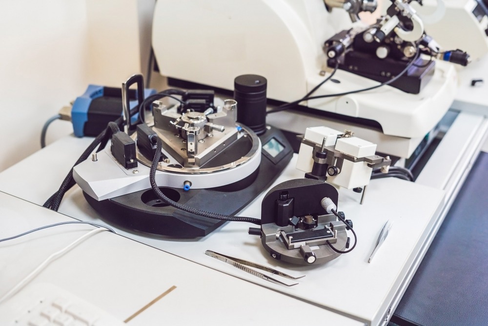
Atomic force microscopy (AFM) was first demonstrated in 1985 by Binnig, Quate and Gerber. Since then, the high-resolution non-optical imaging technique has become a powerful tool for surface analysis.

Image Credit: Elizaveta Galitckaia/Shutterstock.com
AFM is an accurate and non-destructive method of obtaining various measurements, including topographical, electrical, magnetic, chemical, optical, and mechanical measurements of a sample surface. AFM can obtain these measurements in air, liquids, or ultrahigh vacuum, making the technique invaluable to advanced scientific laboratories and helping to advance in numerous science fields.
How Does Atomic Force Microscopy Work?
The methodology of atomic force microscopy (AFM) is analogous to scanning tunneling microscopy, which uses a sharp tip to raster-scan the surface of a sample, generating a feedback loop to make fine adjustments to the parameters required to produce an image of the sample’s surface.
AFM differs from scanning tunneling microscopy as it does not require a conducting sample, rather than using the quantum mechanical effect of tunneling, AFM leverages atomic forces to map out the interactions between sample and tip.
Various AFM methodologies enable almost any measurable force interaction, including electrical, magnetic, thermal, and van der Waals.
Applications of Atomic Force Microscopy
To date, a vast number of applications of AFM have been developed. These applications are helping to further research and development in a wide range of scientific fields.
There are several main benefits of AFM, which have encouraged it to be adopted by a wide range of disciplines, including the capability to image structures at the nanoscale and in 3D, measure samples under near-physiological conditions (e.g., no damage or staining required), obtain data in real-time and obtain structural and mechanical information.
Some of the most common applications of AFM include imaging biological molecules, intracellular environments, cells or tissues, polymers, ceramics, composites, glass, and nanostructures. AFM has been beneficial to the field of cancer research and diagnosis.
Recent Developments of Atomic Force Microscopy
Because AFM enables nanometer-scale investigation of cells and molecules, it has been incredibly beneficial to cancer research. To date, two of the most critical applications of AFM in oncology include diagnosing single cells in cancer, observing changes between cancerous vs. non-cancerous cells, and developing anti-cancer pharmaceuticals.
In 2021, researchers at the University of Sheffield, UK, published findings of their research that had developed a novel method of using AFM to characterize metastatic breast tumors.
The team established a method of quantifying the micro-mechanical properties of breast cancer, such as the elastic moduli and viscosity.
Using AFM, the team took measurements without damaging the breast cancer bone metastases samples or the surrounding microenvironment. Research such as this demonstrates how AFM is crucial in understanding the mechanisms underlying disease development, progression, and metastasis in cancer.
Other recent developments include using AFM to develop our understanding of the SARS-CoV-2 virus responsible for the COVID-19 pandemic.
One study, published in Scientific Reports in June 2021, demonstrated how AFM could effectively analyze single SARS-CoV-2 viruses.
The team’s method used AFM to accurately depict the structurally intact infectious and inactivated virus. Their combination of AFM and plaque assays was fundamental for accelerating research against the COVID-19 pandemic. The technique will likely be leveraged for the study of other viruses also.
Challenges Facing Atomic Force Microscopy
While AFM is a well-established research tool across worldwide laboratories, it is not without its limitations. For example, AFM requires samples to be adsorbed onto supporting surfaces. This can alter the vesicles' size and shape, deforming the sample.
Other challenges include the single scan image size, which is much larger than that of the scanning electron microscope: order of 150 x 150 micrometers vs. millimeters.
AFM has a relatively low scan time compared with similar technologies, which can impact the sample's thermal drift. Finally, the periodic contact of the probing tip can inadvertently drag liposomes across a sample. However, despite this limitation, AFM is still used in evaluating liposomes.
The Future of Atomic Force Microscopy
AFM is already established as a well-trusted technique for the study of a wide range of samples, both biological and non-biological.
However, the method is still yet to reach its full potential. Recent research continues to develop the method for use in novel applications.
In the future, we will likely see the continued development of AFM for use in cancer research, where it is already making a significant impact.
We will also likely see further use of the technique in virology, given its importance in the recent fight against the COVID-19 pandemic.
References and Further Reading
Chen, X. et al. (2021) Atomic Force Microscopy reveals the mechanical properties of breast cancer bone metastases. Nanoscale, 13(43), pp. 18237–18246. https://pubmed.ncbi.nlm.nih.gov/34710206/
Deng, X., Xiong, F., Li, X. et al. (2018) Application of atomic force microscopy in cancer research. J Nanobiotechnol 16, 102. https://doi.org/10.1186/s12951-018-0428-0
Last, J.A. et al. (2010) The applications of Atomic Force Microscopy to vision science. Investigative Opthalmology & Visual Science, 51(12), p. 6083. https://doi.org/10.1167/iovs.10-5470
Lyonnais, S., Hénaut, M., Neyret, A. et al. (2021) Atomic force microscopy analysis of native infectious and inactivated SARS-CoV-2 virions. Sci Rep 11, pp. 11885. https://doi.org/10.1038/s41598-021-91371-4
Tseng, A.A. (2011) Advancements and challenges in development of atomic force microscopy for nanofabrication. Nano Today, 6(5), pp. 493–509. https://doi.org/10.1016/j.nantod.2011.08.003
Disclaimer: The views expressed here are those of the author expressed in their private capacity and do not necessarily represent the views of AZoM.com Limited T/A AZoNetwork the owner and operator of this website. This disclaimer forms part of the Terms and conditions of use of this website.