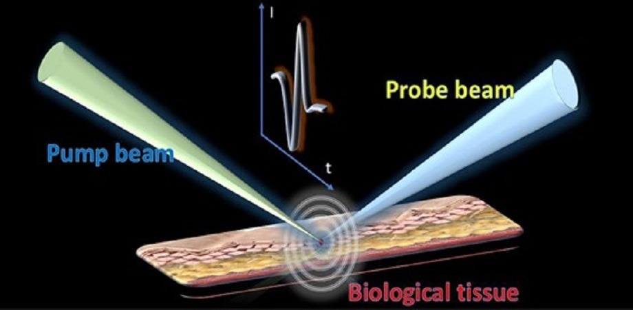
Mechanism of photoacoustic remote sensing. Image Credit: Guyue Hu, PARS Mechanism, 2023
When a pulsed laser sheds light on the surface of biological tissue, part of the photon energy is absorbed by the tissue for heat to be produced. The increase in heat results in the tissue’s thermoelastic expansion, thereby releasing energy in the form of ultrasonic waves. By scanning the sample and gathering the matching photoacoustic signals, scientists are capable of reconstructing 2D or 3D images of the biological tissue.
Generally, ultrasonic transducers gather photoacoustic signals. However, since sound waves attenuate in air, it is usually essential to add ultrasonic gel or water between the tissue sample and the transducer to guarantee signal detection sensitivity.
This physical contact or immersion might have considerable effects on biological samples, thereby greatly restricting the suitability of traditional photoacoustic imaging in several practical scenarios.
Conversely, as a result of the material and innate structural properties of ultrasonic transducers, their central response frequency and detection bandwidth have been restricted, which might decrease the sensitivity of the system concerning broadband signal detection. Taking such limitations into account, conventional photoacoustic imaging needs an update for high-quality photoacoustic research.
Photoacoustic remote sensing imaging is known as a novel photoacoustic imaging modality. Unlike traditional acoustic detection with the help of ultrasonic transducers, photoacoustic remote sensing makes use of another laser beam for acoustic signals to be detected.
Particularly, one more laser source has been adopted as the probe beam that is confocal with the excitation beam. When the energy is absorbed by the sample to produce initial pressure, the sample surface’s refractive index changes as a result of the elasto-optical refractive index modulation.
By tracking the reflection intensity of the probe beam, the matching photoacoustic signals might be examined. All-optical detection of acoustic signals removes direct contact with the sample.
At the same time, as a result of the optical sensing, the detection bandwidth might be transferred merely from the restricted ultrasonic transducer to an extensive photodiode. This offers the chance to additionally enhance the signal-to-noise ratio and detection sensitivity of the system.
Based on the above findings, a research group from the University of Hong Kong has reported near-infrared photoacoustic remote sensing microscopy for noncontact imaging of lipids.
As reported in the journal Advanced Photonics Nexus, the group’s photoacoustic remote sensing microscopy makes use of a 1.7-μm thulium-doped fiber laser as the pump beam to optionally stimulate the C–H bond in lipids.
Simultaneously, another laser measuring around 1.5-μm continuous-wave (CW) has been adopted as the detection beam that is confocal with the pump beam to detect the initial ultrasonic pressure.
The optical detection of ultrasonic signals eliminates the need for ultrasonic transducers and enables remote sensing of photoacoustic signals. This technique provides a wide detection bandwidth and improves the signal-to-noise ratio and detection sensitivity of the system.
In their experiment, the research group demonstrated the imaging results of two types of pure lipid samples using photoacoustic remote sensing. They also analyzed the comparable power spectrum density of the signals.
Compared to conventional transducers, the optical detection technique provided a broader frequency response. Furthermore, the team conducted photoacoustic remote sensing imaging on biological samples, including brain slices and nematodes, which exhibited excellent contrast and signal-to-noise ratios. This demonstrated the high-performance imaging capability of the technique on a tissue scale.
Photoacoustic remote sensing microscopy achieves label-free imaging that can target specific molecular bonds.
Kenneth K. Y. Wong, Study Corresponding Author and Professor of Engineering, University of Hong Kong
Wong added, “Optical detection of ultrasonic signals provides noncontact operation and a broader frequency response. Meanwhile, the photoacoustic remote sensing microscopy shows high performance for lipid distribution mapping on the tissue scale.”
The newly developed method indicates great application potential for a range of biomedical research.
Journal Reference
Hu, G., et al. (2023) Noncontact photoacoustic lipid imaging by remote sensing on first overtone of the C-H bond. Advanced Photonics Nexus. doi.org/10.1117/1.APN.2.2.026011.