Reviewed by Mila PereraSep 23 2022
Scientists have formulated a basic and rapid method to carry out optoretinography, an imaging method that assesses light-stimulated functional activity in the retina of the eye. The retina is a system of neurons in the back of the eyes in charge of sensing light and providing vision.

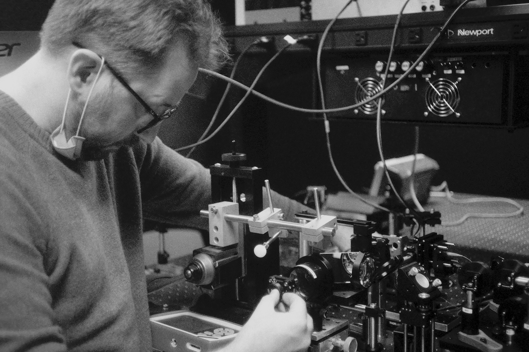
First author Kari Vienola is shown with the velocity-based optoretinography optical setup. The new approach developed by the researchers could help accelerate the development of new treatments for retinal diseases. Image Credit: Ravi Jonnal, University of California, Davis
In the United States, over 50% of people aged 60 and older are afflicted with retinal diseases such as diabetic retinopathy and macular degeneration. These diseases impact the retina’s working in ways that decrease eyesight and can lead to blindness if not medically checked. The new method could accelerate the creation of new treatments for eye ailments.
Optoretinography has typically used very costly equipment that required multiple experts to operate while also producing enormous data volumes requiring extensive computational resources. We devised a way to do it cheaper and quicker.
Ravi Jonnal, Study Lead Author, Vision Science and Advanced Retinal Imaging Laboratory, Department of Ophthalmology and Vision Science, University of California, Davis
Jonnal and contemporaries report their new method, which they refer to as velocity-based optoretinography in Optica, Optica Publishing Group’s journal for challenging research. They also show the ability of the technique to assess retinal response in three healthy study participants.
"Although velocity-based optoretinography could potentially provide clinicians with more accurate and earlier information about functional losses in the retina, its first real impact is more likely to be in expediting clinical trials for new treatments of retinal diseases." Added Jonnal.
He continued, "If we can detect whether retinal function is getting better or worse faster than with traditional tests such as eye charts, it will greatly accelerate the development of treatments."
Jonnal carried out a few of the first optoretinography measurements as a doctoral student in Don Miller’s lab at Indiana University.
Tracking Shape Changes
Optoretinography can sense minor variations in the shape of neurons that conduct or produce signals in the retina.
Jonnal and other researchers have employed adaptive optics and optical coherence tomography (OCT) to visualize and monitor these neurons in the active, moving eye and then employed motion correction algorithms to stabilize the images and derive the functional response.
This expensive and laborious process requires resolving and monitoring the position of each cellular feature and using those positions to establish if the cell's shape has changed.
“When we use one of our adaptive optics systems to make optoretinography measurements, the experiment can easily take half a day and result in a terabyte of data that needs to be processed. Processing the data to extract a functional signal takes, minimally, another day or two,” states Jonnal.
To circumvent the necessity of resolving and monitoring separate neurons, Jonnal and the team were keen to see if they could compute the speed, or velocity, at which the retinal neurons travel relative to each other.
We believed that even if the positions of the features vary from cell to cell, the speed at which they move relative to one another would be highly correlated among cells. This proved to be correct.
Ravi Jonnal, Study Lead Author, Vision Science and Advanced Retinal Imaging Laboratory, Department of Ophthalmology and Vision Science, University of California, Davis
Measuring Moving Neurons
To conduct velocity-based optoretinography, the scientists designed a new OCT camera that enables a single operator to collect images from more areas in the retina than is possible using other methods of optoretinography.
The team demonstrated their new method by applying it to gather measurements from three healthy participants. They were able to obtain data from each patient within 10 minutes and have that data analyzed and results available in about 2 to 3 minutes.
They demonstrated that the functional optoretinographic responses were calculated with the approach scaled with the light stimulus dose and that the dose-stimulus response was reproducible among the volunteers.
The team are currently scheduling experiments to present the method’s sensitivity to disease-associated dysfunction. Jonnal is also partnering with clinicians at the University of California, Davis, to apply it to patient imaging and to help in deducing results from stem cell therapies and gene therapy treatments for hereditary retinal diseases.
The scientists are aiming to also use the new optoretinography method in animal models of retinal disease.
Journal Reference
Vienola, K. V., et al. (2022) Velocity-based optoretinography for clinical applications. Optica. doi.org/10.1364/OPTICA.460835.